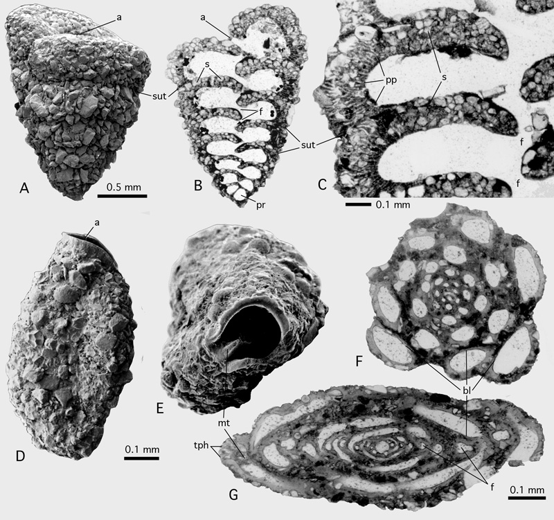
Figure 6: Agglutination in foraminiferan walls; Gulf of Aqaba, Red Sea; Recent.
A-C: Textularia sp. C in et alii, 1993. A: Lateral view showing coarse grains producing a rugged shell surface exept on apertural face. SEM. B: axial thin section of the biserial test showing distribution of agglutinated grains within the shell walls. Light microscope, transparent light. C: Detail from another specimen showing parapore texture of wall and its relation to the agglutinated grains. Transmitted light. D-G: Agglutinella sp., an agglutinating, porcelaneous miliolid. D: lateral view showing coarse agglutination at the shell's suface. E: apertural view showing large aperture with a porcelaneous peristomal rim and a miliolid tooth. SEM.
F-G: Schlumbergerina alveoliniformis (),
thin sections in the - and perpendicular to the - apertural axis, showing early growth stages without apparent agglutination and a very thin basal layer coating the rugged surface of adult chambers of the previous whorl. Transmitted light.
a: aperture; bl: basal layer; f: foramen; mt: miliolid tooth; pp: parapores; pr: proloculus; s: septum; sut: (chamber) suture; tph: trematophore.