The magnification of each figure is in proportion to the length of the 10 cm scale bar above. |
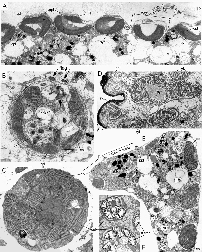
Figure 30: Chloroplasts of foraminiferal symbionts. Gulf of Aqaba. TEM graphs courtesy .
A: Bacillarian (diatom) symbionts below lateral chamber wall, housed in eggholders, below pore mouths, of Amphistegina lobifera .
x 5,000. B: dinophycean (dinoflagellate) symbiont of Amphisorus hemprichii . Note sections of short flagellae (arrows) permitting active movement of the symbiont within the lacunar system of the host, to regulate its access to light. The nucleus with its (polyploid) chromosomes visible during the interphase unusual among eucaryotes but characteristic for dinophyceans. x
10,000. C: rhodophycean (red algal) symbiont of Peneroplis planatus ( et ). x
12,000. D: Bacillarian symbiont of Assilina ammonoides (), ultrathin section oblique-tangential to the lateral surface of achamber. Note the loose stacking of the thylacoids in the chloroplast, a characteristic of symbionts of Assilina. X
8,000. E: Elphidium craticulatum ( et ), ultrathin section through a protoplast of a chamber including one of its retral processes (double arrow), showing free chloroplasts in the host chamberplasm, a characteristic of symbiont husbandry. x
4,000. F: Rhodophycean symbionts of Peneroplis planatus ( et ) from Elba in the Mediterranean. Note starch grains covering the pyrenoid and filling most of the symbiont's mass. The starch grains may also appear as free grains in the host chamber plasm and represent seasonal food storage for the host. x
4,000.
bD: basal pore disc; cpl: chloroplast; flag: flagellum with its base; ilD: interlamellar disc of pore; m: mitochondrium; n: nucleus; OL: organic lining; p: pore; Pl: plasmalemma; ppl: pore plug; pyr: pyrenoid; spl: (mineralized) sieve plate; tyl: thylacoid; v: vacuoles.