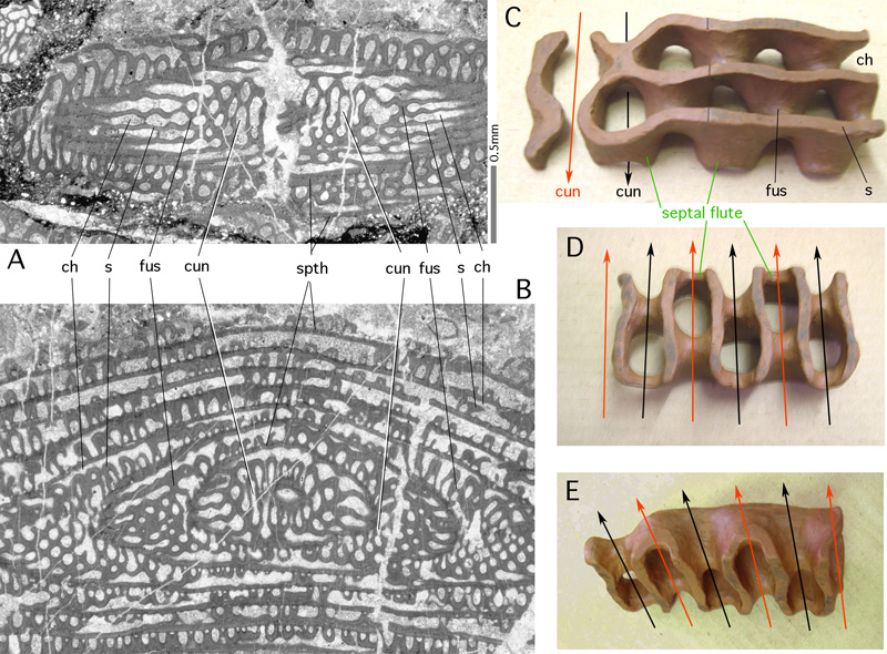
Figure 35: Cuniculi and septal fluting in Eopolydiexodina sp. Transmitted light micrographs. collection, Permian, Iran.
A: shallow tangential section parallel to coiling axis. B: Deep transverse section parallel to coiling axis. C-E: plasticine model sculptured about 1945 by (* 1896 - † 1984). C: Oblique peripheral view showing undivided peripheral parts of two successive chambers. Arrows point in the direction of growth. B: Proximal view showing cuniculi (arrows). C: oblique proximal view showing fluted septal face and cuniculi.
ch: chamber lumen; cun: cuniculus; fus: point of fusion of subsequent septal flutes in opposing positions; s: septum; spth: spirotheca carrying a keriotheca.