The magnification of each figure is in proportion to the length of the 10 cm scale bar above. |
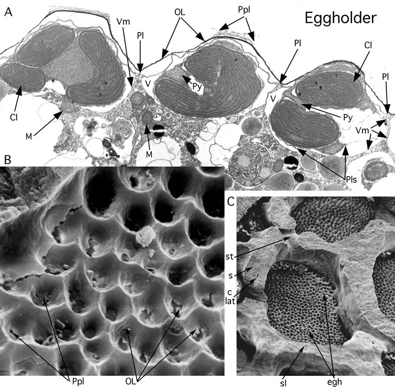
Figure 40: Eggholders harbouring the symbionts in Heterostegina depressa d'. Gulf of Aqaba. Recent.
A: TEM micrograph of section perpendicular to lateral chamber wall; biomineralized wall dissolved, organic cell walls distinctly separated by the techniques of preparation. Courtesy . x
10,200. B-C: dried shell, split open in the equatorial plane. The internal surface of the lateral chamber wall bears eggholders. SEM graphs, x
5,000 and x 500. Drying strechted the organic cell walls.
Cl: chloroplast of bacillariophycean symbiont; clat: lateral intraseptal canal; egh: eggholder; M: mitochondria; OL: organic lining of host; Pl: plasmalemma of host; Plp: pore plug (see pore); Pls: plasmalemma of symbiont; Py: pyrenoid; s: septum; sl: septulum; st: stolon (Y-shaped); V: Vacuole housing symbiont; Vm: vacuolar membrane.