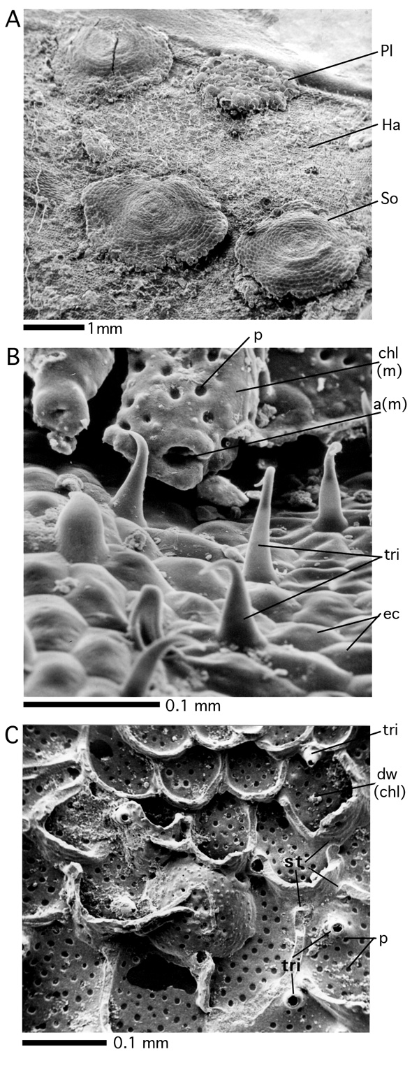
Figure 42: Epiphytic foraminifera and trichomes. Recent. Gulf of Aqaba, Red Sea. SEM graphs.
A: Epiphytes on Halophila leaf. Pl: Planogypsina acervalis (); Ha: surface of Halophila leaf with leaf hairs (trichomes); So:
Sorites orbiculus (). B: Trichome on Halophila leaf and aperture in a radially-marginal position of the epiphytic Planogypsina acervalis (). C: Opened test of Planogypsina acervalis showing the inner side of the perforate, dorsal chamberlet walls, coating the overgrown trichomes of their substrate, and the dorsal part of the septal walls with stolons representing intercameral foramina.
a(m): aperture of a marginal, terminal chamberlet; chl(m): chamberlet of the ultimate marginal chamberlet cycle; dw(chl): dorsal wall of chamberlet; ec: epithelial cells of the Halophila substrate; p: pore; st: stolon; tri: trichome.