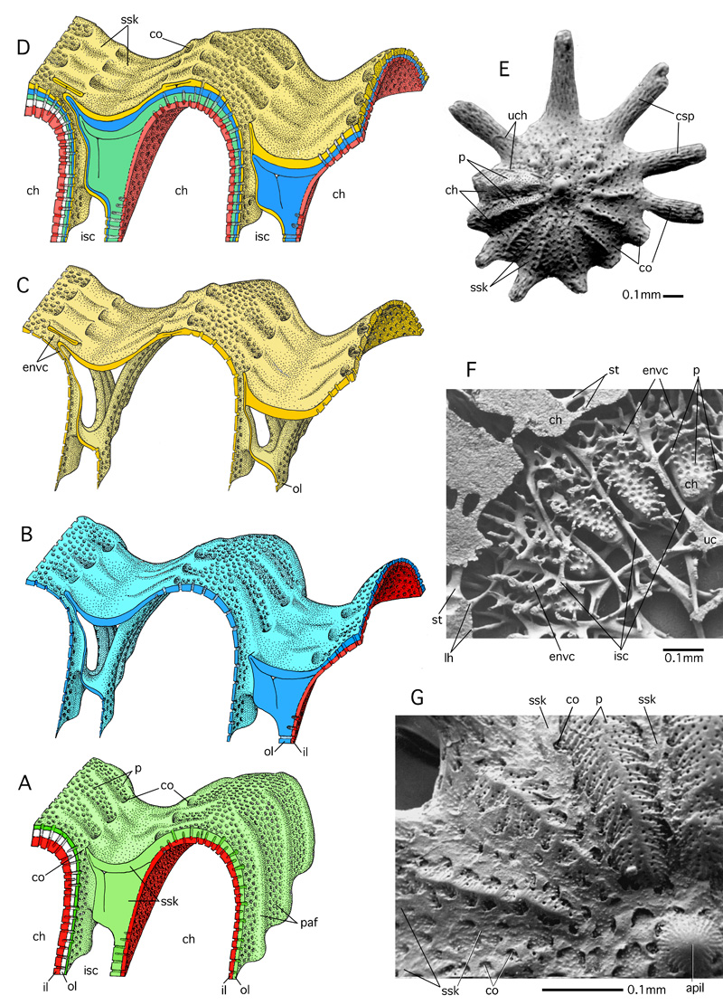
Figure 44: Enveloping canals produced by folded outer lamellas.
A-D: Lamellae in the peripheral portion of three successive chambers. Stereograph, schematic, not to scale. A: ultimate and penultimate chamber: red: inner lamella; green: outer lamella of ultimate chamber, white: outer lamella of penultimate chamber. B: addition of the next chamber with an inner lamella in red and an outer lamella in blue. C: addition of an other chamber with its outer lamella in yellow. D: superposition of lamellas B and C over the then final chamber A after two additional growth steps. E: SEM micrograph of the complete test of Calcarina defrancii d', ventral view, showing distribution of canal orifices over all the test including the canaliferous spines. F: SEM micrograph of an epoxy resin cast of the shell cavities in Calcarina gaudichaudii d' cut in a direction perpendicular to the axis of the spiral shell, ventral view; G: detailed SEM micrograph of the ventral shell surface of C. gaudichaudii. Calcarinas from Keij Island, Indonesia. Recent. After and , 1980.
apil: axial pile of lamellae; ch: chamber and chamber lumen; co: canal orifice; csp: canaliferous spine; envc: enveloping canal; isc: intraseptal space or canal; il: inner lamella; lh: loop-hole; ol: outer lamella; p: pore; paf: perforate part of apertural face; ssk: supplemental skeleton; st: stolon; uc: umbilical canal network; uch: ultimate chamber suture, chamber walls broken off.