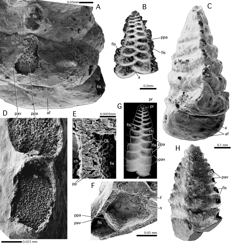
Figure 51: Fistulose chamberlets. All specimens from the Gulf of Aqaba, Red Sea, Recent.
A-C: Sahulia kerimbaensis (). A: Detail of an edge view, SEM graph; B: side view, X-ray graph, black background
removed. Note the narrowness and the irregularity of the fistulose chamberlets in this species. C: edge view, SEM graph. D-H: Spirotextularia floridana (). D: paraporous partition separating the chamber lumen from a fistulose chamberlet. SEM graph. E: Detail of sectioned paraporous partition. SEM graph. Note the organic lining that closes off the parapores from the chamber lumen. SEM graph. F: Shell fragment broken in a direction perpendicular to the shell axis, showing paraporous partition between the chamber lumen and the fistulose chamberlet. SEM graph. G: Lateral view, X-ray graph. H: lateral view of an entire specimen. SEM graph. Note the spiral arrangement of the nepionic chambers.
a: aperture; af: apertural face; ch: chamber lumen; f: foramen; fis: fistulose chamberlet; OL: organic lining; pp: parapore; ppa: paraporous partition; pr: proloculus; pv: pavement; s: septum.