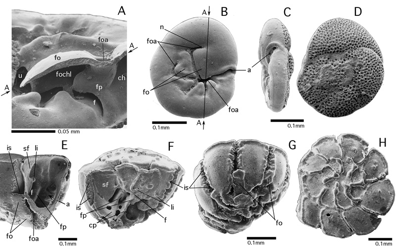
Figure 53: The folium and its apertures. All specimens from the Gulf of Aqaba, Red Sea, Recent. SEM graphs.
A-D: Rosalina bradyi (). A: detail of dissected specimen, oblique-ventral view. The approximate position of the breakage surface (arrows A-A) is indicated by the line A-A in Fig. B. B: ventral view showing the folium
at its maximum development, with anterior and posterior apertures. C-D: peripheral and dorsal views. Note the restriction of the perforation to the dorsal surface of the shell, an indication that the face extends from the umbilical side of the shell over its periphery. E-H: Asterorotalia gaimardi (d'). E-F: dissected specimens showing details of advanced umbilical architecture covered by the folia: foraminal and coverplates (compare
Fig. 34). G: oblique-ventral view showing the folia that cover the ventral part of the interlocular space. H: dorsal view showing the spiral arrangement of the chambers.
a: aperture; ch: chamber lumen; cp: coverplate; f: foramen; fo: folium; foa: foliar aperture; fochl: foliar chamberlet lumen; fp: foraminal plate; is: interlocular space; li: lip (of foramen); n: notch; sf: septal flap; u: umbilicus.