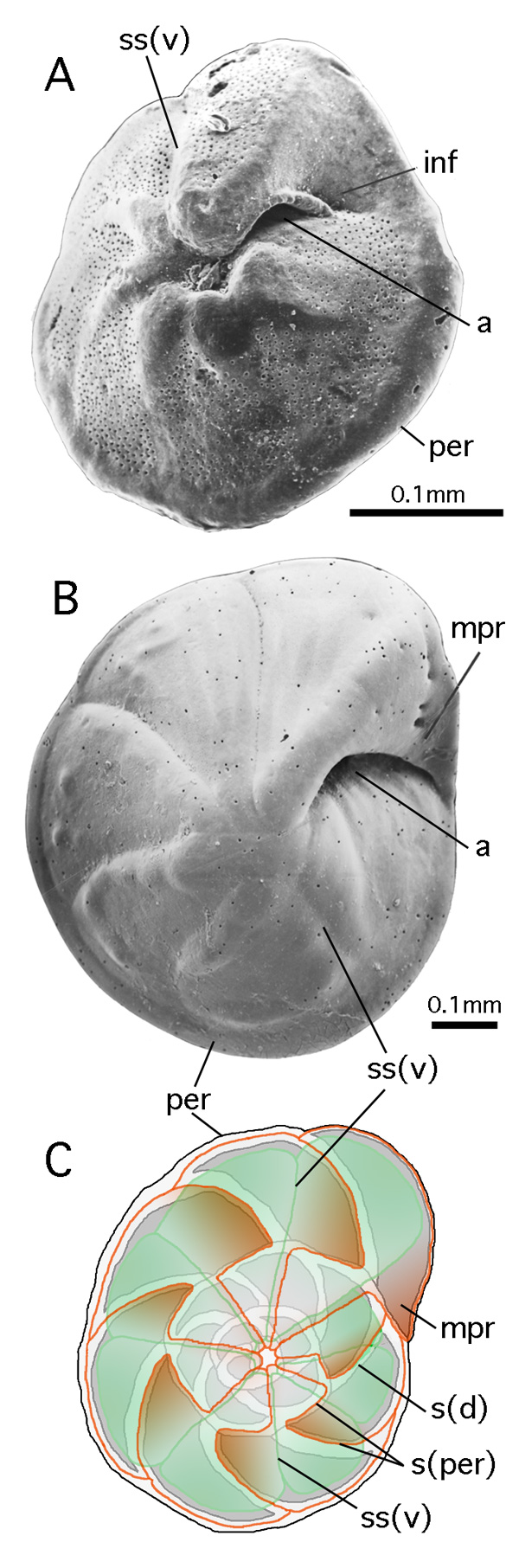
Figure 61: Infundibulum and marginal prolongation.
A: Neoeponides bradyi (), oblique-ventral view showing infundibulum.
SEM graph; Gulf of Aqaba, Red Sea; Recent.
B: Eponides repandus ( et ), ventral view showing marginal prolongation.
SEM graph; Gulf of Aqaba, Red Sea; Recent.
C: Schematic drawing showing position of marginal prolongations in respect to ventral and dorsal test morphology in the last whorl of Eponides repandus.
Gray: dorsal view with raised sutures; red: outline of chamber walls at level ventrally below periphery; green: ventral ouline of chambers below level of marginal prolongation.
a: aperture; inf: infundibulum; mpr: marginal prolongation; per: (imperfotate) periphery; s(d): (oblique) septum (as seen in dorsal view); s(per): position of septum at level of periphery; ss(v): (radial) septal suture in ventral view.