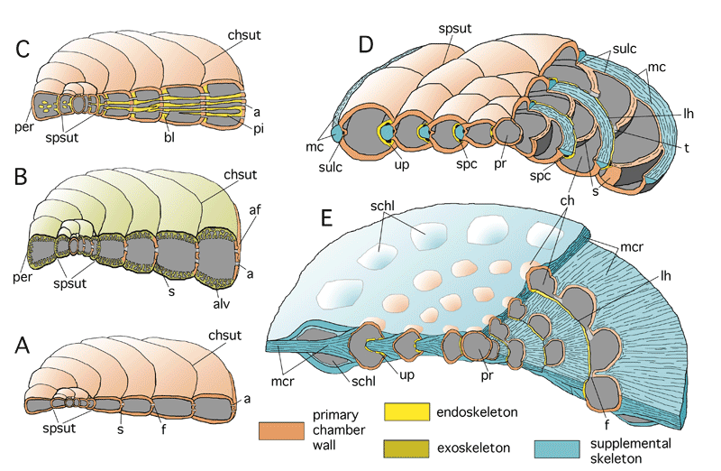
Figure 63: Comparison of foraminiferal skeletons. Schematic, not to scale. Lamellation, perforation and canal orifices omitted.
A: a planispiral-evolute shell without skeletal structures, composed of simple primary chamber walls with multiple apertures, such as
that of Peneroplis. B: a planispiral-evolute shell with an alveolar exoskeleton, such as Pseudocyclammina. C: a planispiral shell
with a pillared endoskeleton such as Archaias. Note that in the axial sections of shells with peneropliform, flaring chambers the periphery of the shell and the
apertural face are on opposite sides. Consequently, the pillars extending from chamber bottom to chamber roof appear in the axial plane
on the side cutting the apertural face as longitudinal and on the other side
cutting the periphery as more or less perpendicular sections. D: a spiral shell with a supplemental skeleton restricted to the periphery of the shell, as in nummulitids
with a marginal cord. E: a spiral shell with an enveloping canal system and a marginal crest as in Pellatispira. Note the primary lateral chamber walls "emerging" from the supplemental skeleton. These primary chamber walls are covered by secondary lamellae but
are perforated in continuation of the primary bilamellar wall. Therefore they
are not a part of the supplemental skeleton. The supplemental chamberlets have perforate lateral walls but do not communicate directly with the spiral chambers by retral stolons. They are fed by canal orifices.
a: aperture; af: apertural face; alv: alveole; bl: basal layer; ch: chamber; chsut: chamber suture; f: foramen; lh: loophole; mc: marginal cord; mcr: marginal crest; per: periphery; pi: pillar; pr: proloculus; s: septum; schl: supplemental chamberlet; spc: spiral canal; spsut: spiral suture; sulc: sulcus; t: tunnel; up: umbilical plate.