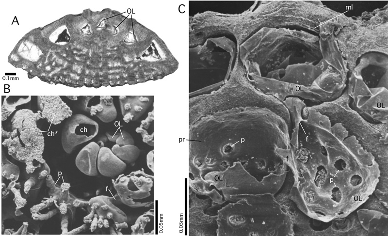
Figure 69: Organic lining.
A: Lockhartia haimei (), megalospheric specimen, axial section, transmitted light micrograph. From the Salt Range, Pakistan. Paleocene. Note the detachment of the organic lining from the inner surface of the biomineralized chamber wall by the first generation of cement deposited during early diagenesis. B-C: Planorbulinella larvata ( et ). Gulf of Aqaba, Red Sea; Recent. B: SEM graph of epoxy resin cast of a microspheric specimen. The subequatorial section of the shell reveals the detail of the early stages of growth, where the resin could not penetrate into the nepionic spiral chambers. After the dissolution of the mineralized shell, the organic lining alone documents the earliest chambers. C: megalospheric specimen, equatorial section, etched in order to reveal the organic lining and the lamellation of the chamber walls. The drying of the preparation before its coating for the SEM contracted the organic lining, detached it from the inner surface of the mineralized wall and broke the connection with the organic poreplugs at their weakest spot. This
produced the circular holes in the organic lining of the preparation. Note the organic lining that coats the foramina connecting the first three chambers.
ch: chamber; ch*: late spiral chamber filled with epoxy resin; f: foramen; ml: median layer of the primary chamber wall separating the inner from all outer lamellae; OL: organic lining; p: pore.