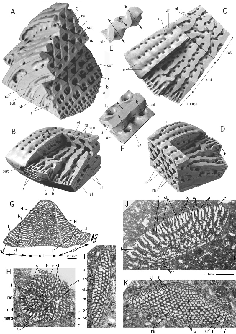
Figure 71: The structure of Orbitolina.
A-D: Oblique and vertical views of the cone base. These plasticine models
sculptured in the years around 1955 by (* 1896 - † 1984) were never published. E-F: Details of a septulum in the radial zone of the discoidal chamber. In model E the septulum is cut below the roof and above the bottom of the chambers. The apparent folding of the section results from the adjustment of the endoskeletal structure to the crosswise-oblique arrangement of the stolon axes (arrows) that produce so-called ramps. In model F the septulum is cut in the middle of the chamber and shows a part of the chamber bottom with the face of the previous chamber. In the middle of the chamber, the section of the septulum appears unfolded. Compare
Fig. 47H and Fig. 80G. G-H: Random thinsections of Orbitolina sp. from Southwestern France, Albian. Transmitted light micrographs. In Fig. G the approximate positions of sections H-K are indicated. Note the inverse orientation: the sections face downward. G-K: Random sections of Orbitolina sp. from Southwestern France, Albian. Transmitted light micrographs. The approximate position of sections H-K are indicated in section G that is very close to the axial plane.
For 's zonation of the chamber see Fig. 20. Section H demonstrates details of the reticular zone, section I details of the marginal zone and section J details of the radial zone. The transverse section K shows the ramps produced by
the crosswise-oblique stolon system.
a: aperture; af: apertural face; b: beam; cl: chamberlet; e: epiderm; f: foramen; gr: coarse grains in the septum that obscure the structural pattern; hor: horizontal section in the plasticine model; marg: marginal zone; r: rafter; ra: ramp; rad: radial zone; ret: reticular zone; s: septum; sf: septal face; sl: septulum; sut: suture of the chambers. Double arrows in E and F: crosswise oblique foraminal axes.