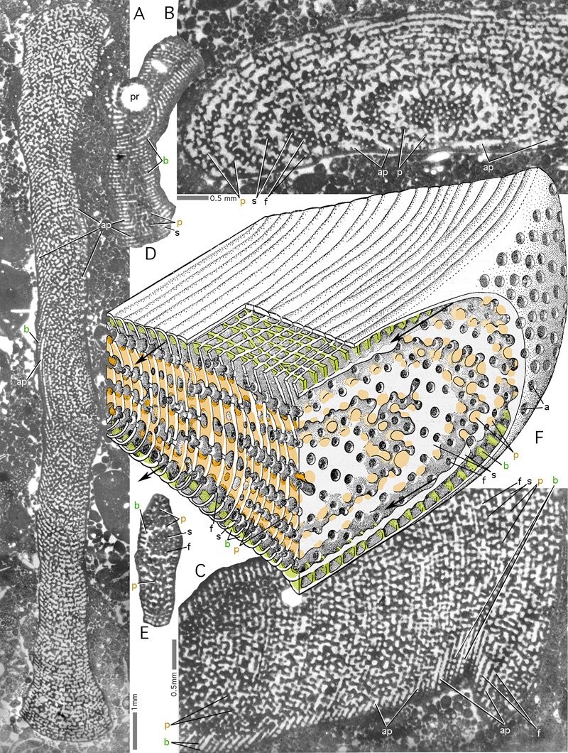
Figure 72: The structure of Orbitopsella:
a simple exoskeleton and a pillared endoskeleton in a discoidal shell: Orbitopsella dubari , Bou Dahar, Eastern Morocco, Middle Lias. Transmitted light micrographs.
A: oblique section of complete microspheric specimen; B: oblique section of microspheric specimen. The septa of the thickened margin are cut tangentially and reveal the alternating pattern
in the disposition of foramina on the septal face. C: oblique tangential section of a microspheric specimen at a low angle to the equatorial plane showing a part of
the disc with its exoskeleton (restricted to beams). Note the large open spaces, the lateral annular passages (arrows), that
separate exoskeleton and endoskeleton. D: Oblique centered section of megalospheric specimen. Note the structured wall of the embryo, that shows it to be a sphaeroconch. E: Transverse section (parallel to
the axis of coiling) of a megalospheric specimen. The septum in this tangential section reveals the alternating pattern of the apertures. F: schematic model of structure after , 1967; not to scale.
Green: exoskeleton; brown: endoskeleton.
a: aperture; ap: annular passage; b: beam; f: foramen; p: pillar; pr: sphaeroconch; s: septum.