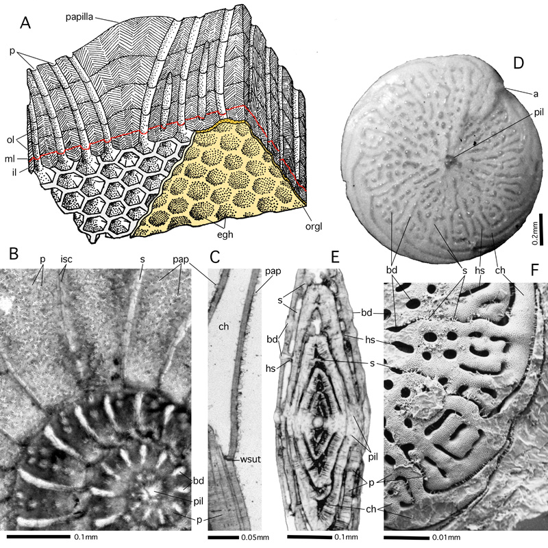
Figure 73: Papillae, beads and axial piles of lamellae.
A: papilla and pores. Stereograph, schematic, not to scale; after , 1977. B-C: Assilina madagascariensis (d') from Mauritius. Recent. Transverse section parallel to equatorial plane (B) and axial section (C) with minute papillae on
the last whorl; transmitted light micrograph. D-F: Beading in Amphistegina papillosa . Gulf of Aqaba, Red Sea; Recent. D: dorsal view of shell showing beading of septular and hemiseptular sutures. Incident light micrograph. E: axial section showing hemiseptular and septular support of beads; transmitted light micrograph.
F: Epoxy resin cast of shell cavity showing spiral main chamber lumina in dorsal view, interrupted by the hemiseptula supporting the interseptal beads, SEM graph.
a: aperture; bd: beads (septular and hemiseptular); ch: main (spiral) chamber lumen; egh: eggholders; hs: hemiseptulum; il: inner lamella; isc: intraseptal canal system; ml: median layer; ol: outer lamellae; orgl: organic lining of protoplast; p: pores; pap: papillae; pil: axial pile of lamellae; s: septum and septal suture; wsut: whorl suture.