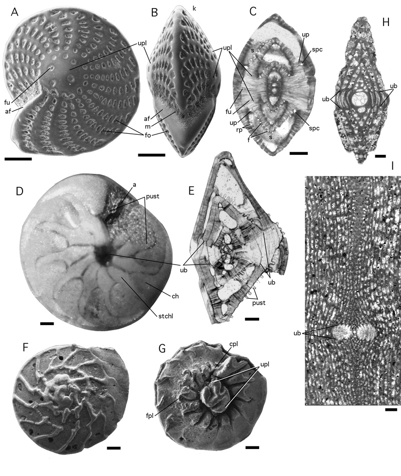
Figure 77: Umbos and umbilical plugs.
A-C: Elphidium craticulatum ( et ) from the Gulf of Aqaba, Red Sea. Recent. A: lateral view, SEM graph. B: peripheral view, SEM graph. C: slightly oblique axial section cutting through the symmetrical pair of umbilical plugs. Note the presence of spaces in the biumbilical shell,
caused by spiral canals and funnels. Transmitted light micrograph. D-E: Amphistegina lessonii d' from the Gulf of Aqaba, Red Sea. Recent. D: ventral view shows the stellar chamberlets overgrowing
part of the ventral umbo. Incident light micrograph. E: axial section of specimen having lost the last few chambers. There is a smaller dorsal and a larger ventral umbo. F-G: Ammonia umbonata (). Dorsal and ventral views, SEM graphs. The ventral (umbilical) view exhibits a free-standing, composite umbilical plug. H-I: the umbos in porcelaneous shells may react to diagenetic processes by
differences in recrystallisation that possibly indicate a differentiation in the texture of the wall in the umbonal area of the shell. Diagnostic for the involute stages
in the growth of Meandropsinidae in the Upper Cretaceous of the Pyrenean Gulf. H: megalospheric Fascispira sp. from Canelles, Lerida, Northern Spain. Axial section. I: microspheric Larrazetia larrazeti (), center of axial section, from Bac de Grillera, Northern Spain. All scale bars 0.1 mm.
a: aperture; af: apertural face; cpl: coverplate; f: foramen; fo: fossette; fpl: foramenal plate; fu: funnel; k: keel; m: mask; pust: pustules on amphisteginid face; rp: retral process; s: septum; spc: spiral canal; ub: umbo; up: umbilical plate; upl: umbilical plug.