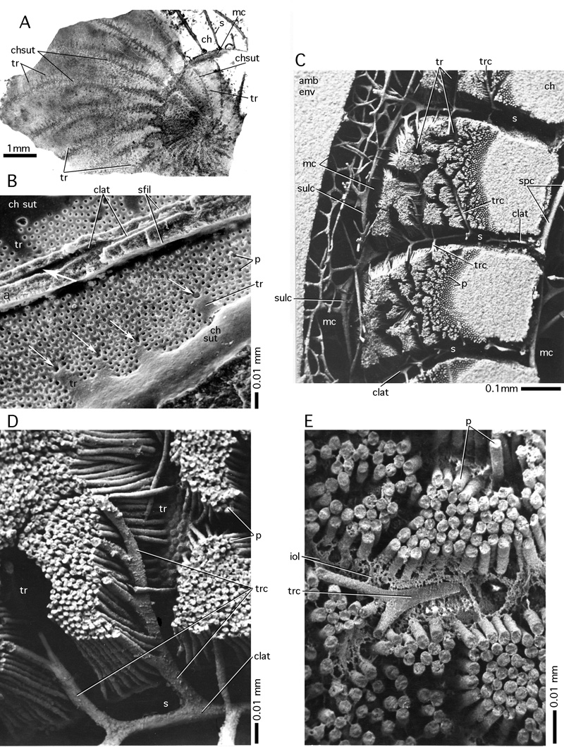
Figure 81: Trabeculae.
A: trabecules in a totally evolute nummulite, Nummulites giganteus (). Crimea. Lower Eocene (Cuisian). Transmitted light micrograph of a section tangential to the lateral chamber walls. B-D:
unfilled shells of Nummulites planulatus () from Bos d'Arros, Gan, Southwestern France, Cuisian. B: lateral surface of the shell with a part of a septal suture between alar prolongations and their extensions, the trabeculae. The white arrows indicate the orifices of the trabecular canals. The lateral surface is overgrown by the next whorl that has been broken away above the basal suture of a septum of an alar prolongation leaving a
linear mark called septal filament. At the base of the septum of an alar prolongation there is a single intraseptal canal running the lenght of the alar prolongation. Where such a canal crosses a trabecular orifice (arrow), a passage may be created by partial resorption of the wall. This passage connects canal cavities of two successive whorls. SEM graph. C: epoxy-resin cast of shell cavities cut in
an approximately equatorial direction showing two complete chambers with their marginal cord. Note the "grooves" in the porous wall created by the trabecular canals. SEM graph. D: detail of C showing the deviation of the pores in order to admit a trabecular canal. E: trabecular canal in Nummulites partschi from Bos d'Arros, Gan. Cuisian. Epoxy resin cast. The preservation of the interlamellar organic lining as
a cavity in the shell permits the oblique path of the trabecular canal to be followed through subsequent lamellae.
Abreviations: amb env: ambient environment appearing in a cast of cavities as solid mass; ch: chamber lumen; chsut: chamber suture; clat: lateral intraseptal canals in the chamber and in the alar prolongations; iol: interlamellar organic layer; mc: marginal cord; p: pores; sfil: septal filament; spc: spiral canal; sulc: sulcus; tr: trabecule; trc: trabecular canal.