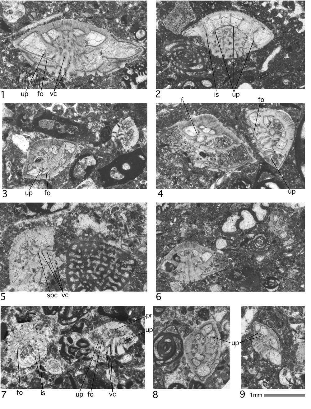
Plate 12, figs. 1-9. Medocia blayensis , 1971.
1: subaxial section of a possibly microspheric specimen. 2: oblique section inclined about 70° to the axis of coiling. It shows the radial arrangement of the vental cameral septa. Note the umbilical plate glued onto the ventral surface of the previous whorl. At the left, an oblique centered section of Penarchaias glynnjonesi.
3: two oblique sections inclined at about 20° and 30° to the axis of coiling.
4: microspheric (left) and megalospheric (right) shells in oblique sections.
5: on the left: a section approximately parallel to a dorsal cone mantel line. On the right: an axial section of the adult part of the cone of Coskinolina cf. liburnica .
6: axial section. On the far right: the axial section of a juvenile specimen of Globoreticulina iranica ,
1978.
7: at the left: Rotaliconus persicus n. gen. n. sp., a section tangential to the convex cone base, and Medocia blayensis , 1971, an axial section.
8: section approximately parallel to the dorsal cone mantel line. In addition, to the left, an oblique section of Austrotrillina eocaenica n. sp.
9: section parallel to the axis of coiling showing the umbilical plates.
Abbreviations: f: foramen; fo: folium; is: intraseptal canal system; pr: proloculus; spc: spiral canal; up: umbilical plate; vc: funnel.