|
Last modified Dec. 27, 2000
FAMILY DIPLOPORACEAE
TRIBE ?
GENUS Spinaporella FLÜGEL et FLÜGEL-KAHLER in
E. FLÜGEL et alii 1984
SPECIES Spinaporella andalusica FLÜGEL et FLÜGEL-KAHLER in
E. FLÜGEL et alii 1984
(by J.C. Braga)
1. Synonymy list
1935b Spongien (?).- Pia
in Schmidt, Pl. II, fig. 3-5
1980 Griphoporella curvata.- Martin & Delgado, Fig.
4
1980 (?) Dasycladacean.- Martin & Delgado, Fig.
6-10
1984 Spinaporella andalusica n. gen. n. sp.- E. Flügel & Flügel-Kahler
in E. Flügel, Flügel-Kahler et alii, Pl. 20; Pl. 21, fig. 1-6 & 8; Pl. 22, fig.
1-9; Pl. 21, fig. 9, from Pia (1935b : Pl. II, fig. 4); Pl. 21, fig. 10,
from Pia (1935b : Pl. II, fig. 3); Pl. 21, fig. 11,
from Pia (1935b : Pl. II, fig. 5)
1987 Spinaporella andalusica.- Braga & Martin, Pl. I, fig. 5 &
10
1989 (?) Dasycladacean.- Martin & Braga, Pl. 2, fig.
1; Pl. 2, fig. 2-3,
from Martin & Delgado (1980 : Fig. 6 & 10)
1999 Spinaporella andalusica.- Parente & Climaco, Pl. 39, fig. 2;
Pl. 40, fig. 1-2 & 7-8
2. Types
Holotype: Pl. 21, fig.
3 [= Pl. 20 pars],
sample 8B, thin
section N° SM.B. 13499, Paläobotanische Abteilung, Forschungsinstitut
Senckenberg, Frankfurt am Main (Germany)
Dimensions (Flügel
& Flügel-Kahler in Flügel et alii, 1984): « Holotype
- outer diameter Ø (D) = 2900 μm, inner diameter Ø (d) = 2300 μm,
d/D = 79,3%, diameter of the first-order branches 30 μm; thickness
of the zone (1) = well-calcified zone near the stem cell - 250 μm.
The holotype is non-fertile. »
Type locality: Pico de la Carne, SSE Monachil - SE La
Zubia, Granada (Spain)
Stratum typicum: Norian. The dolostones of Pico de la Carne section were attributed by Flügel
et alii (1984) to the Middle Triassic. Braga & Martin (1987) demontrated the Late Triassic (Norian) age of these materials.
3. Diagnosis
Original diagnosis
(Flügel
& Flügel-Kahler in Flügel et alii, 1984): « Remarkable long cylindrical thalli
(Pl. 20) with variously calcified walls which may exhibit up to three morphological zones
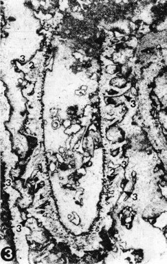
(Pl. 21/1, 21/2, 21/4, 22/3, 22/4); see diagnosis of the genus. Primary branches show a metaspondyle arrangement.
Clusters consist of several thread-like branches which are oriented vertically or slightly obliquely to the main axix in the area adjacent to the stem cell. In the outer part the branches curve upwards and are generally poorly calcified
(Pl. 21/4).
A better calcification is found in the marginal zone of the thalli where the branch bundles are covered by a joint calcareous sheath
(Pl. 21/3).
The fertile stage is characterized by a spinose calcification pattern at the periphery and by an extremely weak or absence of calcification of the stem cell and the interspaces between the stem cell and the periphery
Typical of the peripheral fragile calcification are elements ressembling Prussian spiked helmets arranged in regular whorls, and pierced by thin branches
(Pl. 21/5, 21/8).
The regular inner curvature of the helmets may indicate the existence of egg-shaped reproductive organs developed outside the stem
cell. »
Reconstruction:
Fig. 2 (Flügel
& Flügel-Kahler in Flügel et alii, 1984).
4. Description
Original description
(Flügel
& Flügel-Kahler in Flügel et alii, 1984): « Non-fertile forms are more common than complete fertile stages, but abundant disintegrated elements of the latter are found in most samples.
Large and small thalli occur in both stages. Non-fertile thalli with a well-defined thin pore-bearing zone around the axial cavity are most common in contrast to thalli exhibiting the other modes of calcification.
The outlines of thalli with pores vary considerably. Cross-sections often exhibit a typical pattern characterized by a concentric medial split within the wall
(Pl. 21/9, 22/1),
caused by the three different zones of calcification. »
Most recent description
(Parente & Climaco, 1999): « The calcareous skeleton is cylindrical, very long (up to 30 mm) and sometimes curved. A relatively wide central cavity (d =
1520-2300 μm) is bordered by the well calcified inner part of the skeleton (350-400 μm thick) passing outward into a poorly and irregularly calcified zone consisting of long and thin apophyses of the calcareous skeleton separated longitudinally by micritized or uncalcified furrows (Pl. 40/1-2). These apophyses start nearly perpendicular to the central stem axis but usually curve upward distally. In some specimens the outer part of the skeleton is characterised by long and thin spines, described by FLÜGEL & FLÜGEL-KAHLER
(in FLÜGEL et al., 1984) as resembling Prussian spiked helmets. These spines, perforated by a thin pore, are found arranged in regular whorls or as individual scattered elements (Pl.
39/2). In some specimens (Pl. 40/7-8) we observed curved structures probably corresponding to the distal end of the egg-shaped cavities described by FLÜGEL & FLÜGEL-KAHLER
(in FLÜGEL et al., 1984). The laterals are characterised by a metaspondile arrangement (Pl. 40/1-2). Each cluster is most probably made of 4 laterals. The shape of the laterals is difficult to reconstruct. The well calcified inner part of the skeleton shows thin and irregular pores (p = 50-75 μm). We can only suppose that, as stated by FLÜGEL & FLÜGEL-KAHLER
(in FLÜGEL et al., 1984), the laterals of Spinaporella andalusica were long and thread-like. »
New description: Cylindrical thalli, up to 33 mm long in the studied samples, sometimes curved. External diameter very variable
(950 to 4550 μm, mean 2780 μm) due to the highly varying calcified wall (150 to 900 μm in thickness). Thread-like, primary branches arranged in clusters (2 to 4) and in whorls closely packed. They spread perpendicularly or slightly inclined upwards in relation to the inner surface of the wall and curve upwards at their distal parts. Branch pores, 38 to 75 μm
(mean 59 μm) in diameter at their base, thin slightly outwards.
Wall very irregularly calcified. Calcification is continuous around the stem, poor to absent around the middle part of the branches, and poor and irregular around their distal end giving to the external surface a spiny or scalloped
appearence. This outer part is frequently not preserved attached to the inner
one.
Measurements:
Original
measurements (Flügel
& Flügel-Kahler in Flügel et alii, 1984): « Holotype
- outer diameter (D) = 2900 μm, inner
Ø (d) = 2300 μm,
d/D = 79,3%, diameter of the first-order branches 30 μm;
thickness of the zone (1) = well-calcified zone near the stem cell -
250 μm. The holotype is non-fertile.
The dimensions of some typical thalli are summarized in Table 1.
Isolated helmets at the periphery of th fertile stage have spines
between 600 μm and
23000 μm long (sample 8 B 3). The diameter of the base of the
spine varies between 120 and 580 μm, the diameter of the
egg-shaped cavity between 120 and 290 μm.
Of special interest are the differences in the vertical distance of
the whorls of the non fertile and fertile stages and the significantly
thinner walls of the stem cell of the fertile stages (50-70 μm
vs. 120-600 μm in non-fertile stages. »
|
Non-fertile stages
|
|
Longitudinal sections
|
|
|
L
|
D
|
d
|
d/D%
|
s1
|
|
h
|
p
|
|
|
|
20000
|
2600
|
1600
|
61
|
300-600
|
|
|
25-50
|
|
|
|
27000
|
3600
|
2400
|
67
|
300-500
|
|
|
25-50
|
|
|
|
29000
|
3000
|
2400
|
80
|
300-500
|
|
140-160
|
30-70
|
|
|
|
22000
|
2200
|
1600
|
73
|
120-350
|
|
50-80
|
25-120
|
|
|
|
|
2500
|
1900
|
76
|
220
|
|
60-130
|
40-80
|
|
|
|
16000
|
2600
|
1700
|
65
|
370
|
|
|
50-80
|
|
|
|
|
2500
|
1300
|
52
|
|
|
120-150
|
30-60
|
|
|
|
|
|
|
3200
|
2500
|
78
|
270
|
|
180
|
40-70
|
|
|
|
|
3000
|
2400
|
80
|
|
|
|
70-90
|
|
|
|
|
2800
|
2100
|
75
|
260
|
|
|
50
|
|
|
|
|
2600
|
2100
|
81
|
|
|
|
25
|
|
|
|
|
2500
|
1900
|
76
|
300
|
|
|
|
|
|
|
|
2400
|
1500
|
62
|
|
|
|
25-75
|
|
|
Fertile stages
|
|
|
L
|
D
(without spine)
|
D
(inner diameter)
|
d/D%
|
s (without spine)
|
basal diameter of the helmets
|
h
|
|
Diameter
of the stem cell
|
|
|
10000
|
2300
|
1800
|
78
|
220-320
|
|
330-400
|
|
600
|
|
|
|
2300-2600
|
1700-2100
|
74-81
|
|
280-620,
mostly 450
|
290-610
|
|
|
|
|
|
1800
|
1600
|
89
|
|
280
|
360-480
|
|
600
|
|
|
|
4300
|
|
|
|
320-480
|
|
|
|
5. Remarks
Other types of specimens occur together with the plants described above. They show a
discontinuously calcified wall made up of spiny bodies with a hemispherical cavity at their base. These bodies are crossed by thread-like pores. These were interpreted by Flügel
& Flügel-Kahler (1984) as fertile stages of the species in which calcification developed only on the outer surface of cladosporic reproductive organs and around the distal end of thread-like branches. After examination of hundreds of examples of the species, only in a few cases a transition between both types of thalli is recognizable with some doubt. This, together with the wide range in size of thalli made up only of spiny bodies, seems to support that both types of thalli are independant and not sequential stages in the development of a single plant.
Cladosporic plants grew as individual thalli in most cases.
6. Stratigraphic range
TRIAS (Pia
in Schmidt ,
1935b), MIDDLE TRIASSIC, ? Ladinian (E. Flügel & Flügel-Kahler
in E. Flügel, Flügel-Kahler et alii, 1984) ,
UPPER TRIASSIC (? Martin &
Braga, 1989) ,
Norian (Martin &
Delgado, 1980; Braga & Martin, 1987; ? Martin & Braga, 1989; Parente &
Climaco, 1999)
The dolostones of Pico de la Carne section were attributed by
E. Flügel
et alii (1984) to the Middle Triassic. Braga & Martin (1987) demontrated the Late Triassic (Norian) age of these materials.
7. Real distribution
Spain
(Pia
in Schmidt, 1935b; Martin & Delgado, 1980; E. Flügel & Flügel-Kahler
in E. Flügel, Flügel-Kahler et alii, 1984; Braga & Martin,
1987; ? Martin & Braga, 1989), Italy
(Parente & Climaco, 1999)
8. Paleoecology
Up to recently Spinaporella andalusica
was only known from the Upper Triassic dolostones of the Alpujarride Complex in Sierra Nevada (Betic Cordillera, southern Spain) - Cahorros de Monachil, Pico de la Carne, Picos de las Dehesillas and Picacho Alto sections.
There it occurs as monocultures, monospecific accumulations, or associated to
Physoporella leptotheca Kochansky-Devide, Gyroporella plumosa (Zanin-Buri) Braga,
Griphoporella curvata (Gümbel) Pia and Spinaporella granadaensis Flügel
et Flügel-Kahler in lagoon (inner platform) deposits.
|