Br@chNet
Obolus

Sous-embranchement (subphylum) des Linguliformea
Classe des Lingulata
Ordre des Lingulida
Super-famille des Linguloidea
Famille des Obolidae
Sous-famille des Obolinae


Genus Obolus  Eichwald, 1829


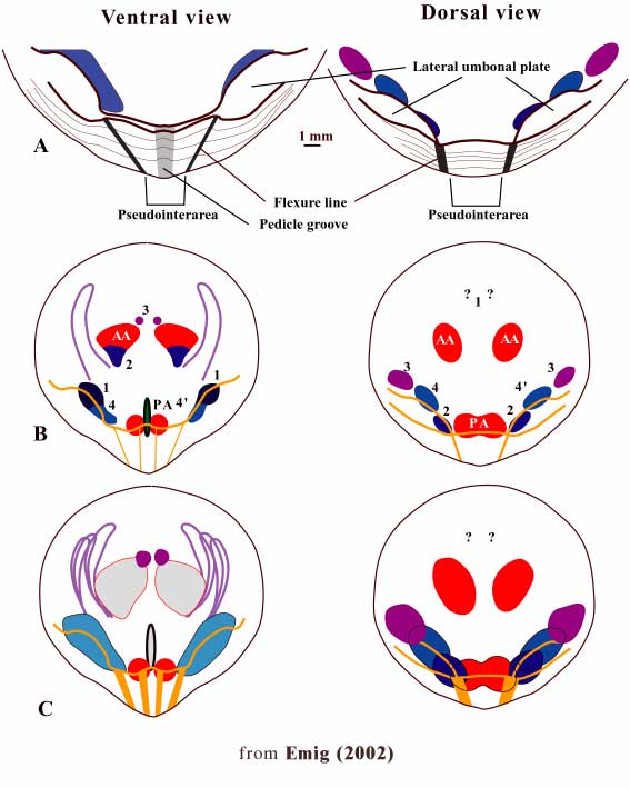
Previous diagnosis in the Treatise (2000):

Shell circular to rounded triangular, dorsibiconvex to subequally biconvex; ventral propareas with deep, narrow pedicle groove; dorsal pseudointerarea lacking flexure lines; visceral area of both valves weakly thickened, extending to midvalve; dorsal median ridge vestigial or absent; vascula lateralia of both valves submarginal, arcuate.

[O. apollinis; SD Davidson, 1853, p. 135; =Obulus Quenstedt,1868, p. 732]

New diagnosis (in Emig, 2002 - see reference below)

Bi-symmetrical muscle arrangement (*)

Ventral valve:
Triangular umbonal region; the pseudointerarea reduced, slightly concave, with elevated flexure lines. Lateral umbonal plates, overhanging the internal side. Posterior adductor muscle paired, separated by short median septum.

Dorsal valve:
Rounded umbonal region with reduced, slightly concave, pseudointerarea with elevated flexure lines. Lateral umbonal plates flat and large overhanging the internal surface. Posterior adductor muscle unpaired.

Middle Cambrian-Ordovician (Tremadoc)

(*) Nota : The Posterior Internal Oblique muscle (numbered 4": Emig 1982) is at present only known in Lingula, Glottidia and Lingularia, which have an asymmetrical muscle arrangement. This feature is probably for the family diagnosis (see above)

  • Emig C. C., 2002. Tools for linguloid taxonomy: the genus Obolus (Brachiopoda) as an example. Carnets de Géologie / Notebooks on Geology, Article 2002/01 (CG2002_A01_CCE)   &  ;[pdf]

  • Emig C. C., 2003. Reply to L.E. Popov and L.E. Holmer (2003): Obolid taxonomy. Carnets de Géologie/Notebooks on Geology, Article 2003/06_Reply (CG2003_A06_Reply_CCE)  &  [pdf]

  • Species of Obolus
    Obolus apollinis Eichwald, 1829

    Synonyms:
    Obolus ruchini Khazanovitch and Popov, 1984
    Obolus transversus (Pander, 1830)
    Obolus rebrovi Khazanovitch and Popov, 1984
    Ungula convexa Pander, 1830

    New Diagnosis (Emig, 2002) :

    including the figure (see below)

    Muscles scars deeply impressed, in particular the postero-lateral ones.

    Ventral valve
    Umbonal region with pseudointerarea forming a beak; reduced subtriangular pseudointerarea, slightly concave, with laterally elevated flexure lines and medially a narrow subparallel pedicle groove. Pseudointerarea generally slightly overhanging the visceral area, but pedicle groove continuous with the internal valve surface.
    Lateral umbonal plates overhanging the internal surface until about the middle of the Anterior Oblique muscle.
    Anterior Oblique and Internal Oblique muscles more or less aligned with the flexure lines of the pseudointerarea.

    Dorsal valve
    Rounded umbonal region; pseudointerarea reduced, triangular, slightly concave, with elevated flexure lines.
    Lateral umbonal plates overhanging the internal side until about the middle of the Internal Oblique muscle.
    More or less extended median ridge at the level of the anterior oblique muscle scars..
    Anterior adductor muscles subparallel to slightly convergent posteriorly.
    Median Lateral Oblique muscle, Anterior Internal Oblique (or Median Internal Oblique) muscle and Anterior Lateral Oblique muscle separated and placed in a line.
    Vascula lateralia on both sides arcuate, peripherally placed (submarginal). Vascula media (dorsal) - no data.


    NOTA : this figure has to be included in the diagnosis because considered as part of this diagnosis.