Br@chNet
Schmidtites

Sous-embranchement (subphylum) des Linguliformea
Classe des Lingulata
Ordre des Lingulida
Super-famille des Linguloidea
Famille des Obolidae
Sous-famille des Obolinae


Genus Schmiditites Schuchert & LeVene, 1929

nom. subst. pro Schmidtia Volborth, 1869 (non Balsamo-Crivelli, 1863)
Schmidtia: Marshall, 1873
Schmidtia: Dall, 1877
Schmidtia: Hall & Clarke, 1892
Obolus (Schmidtia): Mickwitz, 1896
Obolus (Schmidtia): Walcott, 1908
Obolus (Schmidtia): Walcott, 1912
Schmidtites: nom. subst. Schuchert & Levene, 1929
Schmidtites: Rowell, 1965
Obolus (Schmidtites): Goryansky, 1969
1989 Schmidtites: Popov & Khazanovitch, p. 114.
Schmidtites: Puura & Holmer, 1993
Schmidtites: Puura, 1996
Schmidtites: Bednarczyk, 1998
Schmidtites: Holmer & Popov, 2000

Previous diagnosis in the Treatise of Invertebrate Paleontology (2000)
[Schmidtites celatus (Volborth, 1869), originally described under the name Schmidtia celata Volborth, 1869, p. 732]

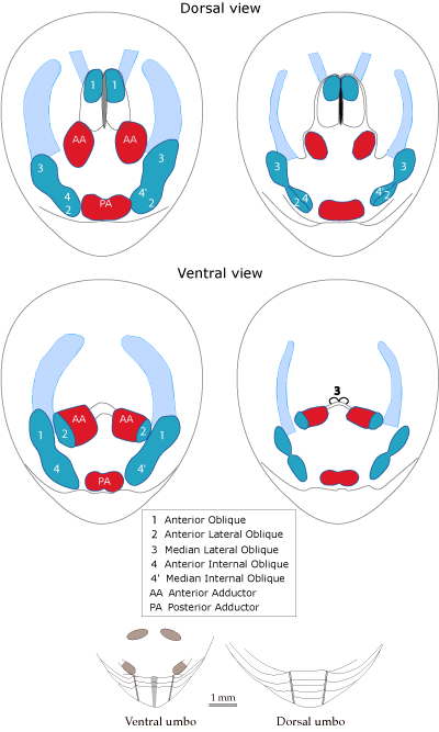
New diagnosis (in Emig, 2006 - see reference below)

Bi-symmetrical muscle arrangement (*)

Ventral valve:
Triangular umbonal region; pseudointerarea slightly concave with well-defined and slightly elevated flexure lines, and with a median narrow pedicle groove.
Posterior adductor muscle unpaired.

Dorsal valve:
Rounded umbonal region; pseudointerarea with well-defined and elevated flexure lines.
Posterior adductor muscle unpaired.
Vascula media short, widely divergent.

Vascula lateralia (main mantle canals) of both sides subparallel to slightly curved.

Shell to subcircular to oval in outline. External surface smooth, with fine concentric growth.

Upper Cambrian and Lower Ordovician (Tremadoc)

(*) Nota : The Posterior Internal Oblique muscle (numbered 4": Emig 1982) is at present only known in Lingula, Glottidia and Lingularia, which have an asymmetrical muscle arrangement. This feature is probably for the family diagnosis (see above)

  • Emig C. C., 2006. Schmidtites celatus (Obolida, Brachiopoda) from the "Obolus sands" (Upper Cambrian - Lower Ordovician) of Estonia. Carnets de Géologie/Notebooks on Geology, Article 2006/04 (CG2006_A04)   [pdf].
  • Species of Schmidtites
    Schmidtites celatus (Volborth, 1869)

    Synonyms:

    Schmidtia celata n. sp.: Volborth, 1869
    Schmidtia celata: Hall & Clarke, 1892
    Obolus celatus: Mickwitz, 1896
    Obolus celatus var. orbiculatus n. var.: Mickwitz, 1896
    Obolus celatus var. praecisus n. var.: Mickwitz, 1896
    Obolus acuminatus n. sp.: Mickwitz, 1896
    Obolus acuminatus var. alatus n. var.: Mickwitz, 1896
    Obolus acuminatus var. humeratus n. var.: Mickwitz, 1896
    Obolus acuminatus var. subtriangularis n. var.: Mickwitz, 1896
    Obolus crassus n. sp.: Mickwitz, 1896
    Obolus crassus var. angulatus n. var.: Mickwitz, 1896
    Obolus (Lingulella) celatus: Walcott, 1898
    Obolus (Schmidtia) celatus: Walcott, 1912
    Obolus (Schmidtites) celatus: Goryansky, 1969

    New Diagnosis (Emig, 2006)

    including the figure A (see below)

    Ventral valve
    Umbonal region with pseudointerarea forming a rounded beak, continuous with the posterolateral margins;
    Pseudointerarea limited laterally by well expressed and slightly elevated flexure lines and separated by a narrow subparallel pedicle groove; width about the half of the internal umbonal portion.
    Muscle arrangement as on figure A.

    Dorsal valve
    Pseudointerarea, slightly concave, width about one third of the umbonal region; limited laterally by well expressed and slightly elevated flexure lines.
    A median ridge extending over 0.6 to 1.5 millimetres at the level of the Anterior Oblique muscle scars.
    Muscle arrangement as on figure A.
    Anterior Adductor muscles slightly divergent anteriorly; the Anterior Internal Oblique (4) (and Median Internal Oblique, 4´) muscle and the Anterior Lateral Oblique muscle (2) form a composite muscle (2 + 4/4´) which is aligned with the close Median Lateral Oblique muscle (3).
    Vascula media short, widely divergent.

    Vascula lateralia, main mantle canals, are subparallel to slightly curved with a similar distance between the tip of the vascula lateralia to the anterior margin of the valve, on both ventral and dorsal sides.

    Shell ventri-biconvex, sometimes thick-shelled: the ventral valve is slightly higher than the dorsal valve, smooth with some concentric growth lines.

    Figure A

    Figure A: Muscle arrangement and disposition of the main mantle canals (their variability, left; in a specimen, right) and umbonal regions (internal view) in Schmidtites celatus (from Emig, 2006).

    NOTA : this figure A has to be included in the diagnosis because considered as part of this diagnosis.


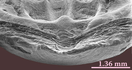
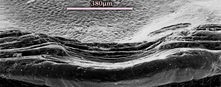
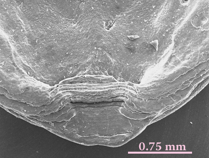
    Umbonal region of ventral valves (internal view)


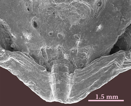
    Umbonal region of dorsal valves (internal view)