Contents
[I - Introduction] [II - Summary of the fraud]
[III - Discussion] [IV- Conclusion] and ...
[Bibliographic references]
Département des Sciences de la Terre, UMR 6538 Domaines Océaniques, Université de Bretagne Occidentale (UBO), 6, avenue Le Gorgeu, F-29238 Brest Cedex 3 (France)
Laboratoire de Paléontologie (C.C.62), Université Montpellier 2, Place Eugène Bataillon, F-34095 Montpellier Cedex 05 (France)
2719 Tyler Street, Long Beach, California 90810 (U.S.A.)
Babeş-Bolyai University, Department of Geology, Str. M. Kogalniceanu nr. 1, 400084 Cluj-Napoca (Romania)
Institut für Paläontologie, Universität Erlangen, Loewenichstr. 28, 91054 Erlangen (Germany)
Manuscript online since May 18, 2009
Starting in 1996 and for almost a decade, M.M. contributed to twelve papers published in international geological journals. These papers dealt with the micropaleontology and biostratigraphy of Cretaceous to Miocene series from Egypt and Libya. They were abundantly illustrated in order to support the author's findings and interpretations. However most photographic illustrations (189 at least) were fabricated with material lifted from the publications of other authors, commonly from localities or stratigraphic intervals other than those indicated by M.M. .
Foraminifera; Corallinales; Dasycladales; Charophyta; fraud; Egypt; Libya.
B., M., E., I.I. & B. (2009).- The case. Additional investigation of a micropaleontological fraud.- Carnets de Géologie / Notebooks on Geology, Brest, Article 2009/04 (CG2009_A04)
L'affaire . Compléments d'enquête sur une fraude micropaléontologique.- À partir de 1996 et pendant près d'une décennie, M.M. a contribué à douze articles parus dans des revues géologiques internationales. Ces publications traitent de la micropaléontologie et de la biostratigraphie de séries d'âge Crétacé à Miocène d'Égypte et de Libye. L'iconographie abondante était sensée renforcer la validité des découvertes et interprétations de l'auteur. Or la plupart des illustrations photographiques (189 au moins) ont été fabriquées à partir de photos "empruntées" à des publications d'autres auteurs, le plus souvent provenant de localités ou d'intervalles stratigraphiques autres que ceux indiqués par M.M. .
Foraminifères ; Corallinales ; Dasycladales ; Charophytes ; fraude ; Égypte ; Libye.
In the period from 1996 to 2003 before (2004) made the initial report on the matter, Mo(u)stafa Mansour published ten papers either alone or as senior author (, 1996a, 1996b, 1998, 1999, 2000, 2001, 2002, 2003; & , 2000; & , 2000), and two papers as junior author ( et alii, 1997; & , 1999). The fraudulent nature of three papers (, 1996a, 2003; & , 2000) has been given wide publicity (, 2004; , 2004a, 2004b; et alii, 2004; et alii, 2008) in the hope of generally deterring such misguided efforts. In order to provide additional support to this inquiry we have undertaken research on the subjects purportly "investigated" (stratigraphy of North Africa, Near East and Middle East and pertinent microfossils). Our intention is to verify all of the descriptions and stratigraphic ages he assigned his figured specimens in order to substantiate more firmly the probability that his findings are unsupported by any valid data. So far we have found 167 more pirated images to add to the 22 discovered by (2004). Four of these twelve papers (, 1999, 2001; et alii, 1997; & , 1999) were published in the Journal of African Earth Sciences and the details of the fraud there were recently exposed in a paper published in that journal ( et alii, 2008). Setting aside the 97 images listed and correlated in that article, 70 remain. As part of a summation of the entire investigation they are discussed in the section that follows.
Earlier in his career was the third
author of a paper in the Neues Jahrbuch für Geologie und Paläontologie,
Abhandlungen ( et alii, 1988).
Following this first promising
publication, (1996a) possibly felt confident enough to submit (in
September 1994) a
manuscript to the same journal and to get it published. This paper deals with
Coralline (red) algae collected in the Middle Miocene strata of Gebel Gushia
(Sinai, Egypt). Surprisingly, the caption for his Fig. 3 (a set of 8
photomicrographs) states aberrantly that the illustrated material is of Middle Eocene
(sic)
age while the legend of his Fig. 4 (a set of 9 photomicrographs) states that the illustrated material is Middle Miocene.
(2004)
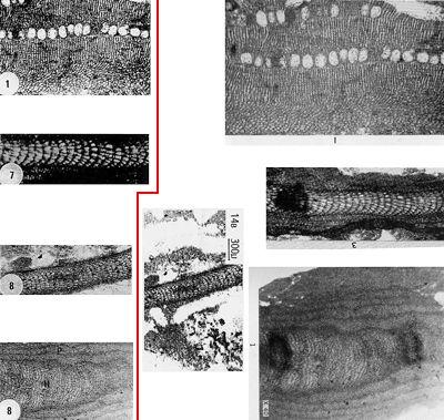
demonstrated that photomicrograph 3.1 labelled "Archaeolithothamnium
saipanense" (Fig. 1 top ![]() ) was reproduced either from
(1957: Pl. 37, fig. 10,
where it was called "Lithothamnium sp." or from his 1961: Pl. 2, fig.
1, "Archaeolithothamnium". Note: we found that himself used
this photo again in 1963: Pl. 25, fig. 3, here again titled "Archaeolithothamnium")
and that it was pirated twice more in &
(2000: Fig. 7.5) and in
(2003: Pl. 3, fig. 1). In addition (Fig. 1
) was reproduced either from
(1957: Pl. 37, fig. 10,
where it was called "Lithothamnium sp." or from his 1961: Pl. 2, fig.
1, "Archaeolithothamnium". Note: we found that himself used
this photo again in 1963: Pl. 25, fig. 3, here again titled "Archaeolithothamnium")
and that it was pirated twice more in &
(2000: Fig. 7.5) and in
(2003: Pl. 3, fig. 1). In addition (Fig. 1 ![]() ), we verified that:
), we verified that:
photomicrograph 3.7 labelled "Lithophyllum prelichenoides" was copied but rotated 180° either from & (1949: Pl. 38, fig. 4, "Lithophyllum aff. prelichenoides") or from (1961: Pl. 10, fig. 3, "Lithophyllum" or 1971: Plate 92, fig. 3, "Lithophyllum");
photomicrograph 3.8 labelled "Jania guamensis" was misappropriated from et alii (1988: Fig. 14B, "Lithophyllum sp.") and rotated 90° clockwise when published;
photomicrograph 4.8 labelled "Lithophyllum kladosum" was taken from (1954: Pl. 192, fig. 1, "Lithothamnium kladosum n.sp.").
Click on thumbnail to enlarge the image.
Figure 1: Left side: 4 images duplicated by (1996a); right side: original images from (1954, 1961) and et alii (1988). [Some rights reserved]
In the same year, (1996b) published in the allied journal Neues Jahrbuch für Geologie und
Paläontologie, Monatshefte another paper, the manuscript of which
was submitted in October 1995. This time he discusses the occurrence of Dasycladalean
(green) algae in the Upper Cretaceous strata of Jebel Um Heriba, Sinai, Egypt.
His Figure 3 (Fig. 2 ![]() ) consists of a set of 8 photomicrographs. We found that all but two of
these images were "borrowed" from
(1991, 1992). The results of
our investigation are summarized in this table:
) consists of a set of 8 photomicrographs. We found that all but two of
these images were "borrowed" from
(1991, 1992). The results of
our investigation are summarized in this table:
| , 1996b | 2006 | et alii, 2006 | et alii, 2006 | ||||
| 3.1 a | Cylindroporella parva | it is not "parva" | 1992 | Pl. III, fig. 2 | Salpingoporella ubaiydhi | it is "ubaiydhi" | |
| 3.1 b | Cylindroporella parva | it is "parva" | 1991 | Pl. 1, fig. 7 | Heteroporella jaffrezoi | ||
| 3.2 a | Heteroporella lemmensis | 1992 | Pl. II, fig. 8 | Dissocladella sp. (it is more probably Cymopolia sp.) | |||
| 3.2 b | Heteroporella lemmensis | 1991 | Pl. 2, fig. 6 | Salpingoporella annulata | questionable "annulata" | ||
| 3.3 | Salpingoporella annulata | it is not "annulata" | 1991 | Pl. 2, fig. 7 | Salpingoporella annulata | questionable "annulata" | |
| 3.5 | Salpingoporella ubaiydhi | it is "ubaiydhi" | 1992 | Pl. III, fig. 4 | Salpingoporella ubaiydhi | it is "ubaiydhi" | |
| 3.6 | Trinocladus tripolitanus | 1992 | Pl. III, fig. 3 | Salpingoporella ubaiydhi | it is "ubaiydhi" | ||
The remaining 2 photomicrographs (Fig. 2 ![]() ) were extracted from E. (1979):
) were extracted from E. (1979):
In a letter to the editors of the Revista Española de Micropaleontología,
said "he used other people's photos because he lacked the means to
provide good illustrations for his manuscripts" (,
2004b). However,
this statement is untrue because his images of Dasycladales (Fig. 2 ![]() )
were deliberately
altered to conceal their adoption much in the way a stolen car is repainted
to hide evidence of the crime.
)
were deliberately
altered to conceal their adoption much in the way a stolen car is repainted
to hide evidence of the crime.

Click on thumbnail to enlarge the image.
Figure 2: Left side: Figure 3 of (1996b); right side: original images from (1991, 1992) and E. (1979). [Some rights reserved]
He was also the second author of a multi-authored paper dealing
with planktonic foraminifera, the manuscript of which was submitted in April
1996 to the Journal of African Earth Sciences
and published the next year ( et alii,
1997). Some of the
photomicrographs of et alii (1988) were
re-used there, but as valid
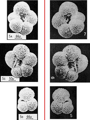
reproductions, for both papers investigate the same locality. However the figures 5
to 7 of Plate 1 (that is Figs. 4.5 to 4.7) of et alii
(1997) are mirror views of the
original photomicrographs in et alii
(1988, respectively Figs. 5D, 5C and 5A): left-coiling planktonic foraminifera
are converted into right-coiling ones, and vice-versa (Fig. 3 ![]() ).
).
Click on thumbnail to enlarge the image.
Figure 3: Left side: original images from et alii (1988 as 5A- Globigerina ciperoensis, 5C- G. angustiumbilicata, and 5D- G. builloides); right side: 3 photomicrographs of et alii (1997 as Globigerina ciperoensis ciperoensis, G. ciperoensis angustiumbilicata, and G. eamesi). [Some rights reserved]
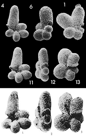
1998 marks 's return to the Neues Jahrbuch für Geologie und Paläontologie, Monatshefte with a manuscript submitted in May 1997. This article deals with the description of a new species of planktonic foraminifer: Clavatorella salumensis n.sp. But his Figure 3.1, called the "holotype", is in fact the holotype of Protentella (Clavatorella) nicobarensis et , 1974 (op. cit.: Pl. 3, fig. 11), which these authors also illustrated in 1975: Pl. 3, fig. 11. 's Figures 3.2 and 3.3, his "paratypes", were also taken from the same paper ( & , 1975: Pl. 3, respectively figs. 12 and 13, a paratype and a topotype of their species). On the basis of an excerpt from 's text: "The new species also shows a great resemblance to Clavatorella nicobarensis & but differs in that the latter taxon (Clavatorella nicobarensis) has less swollen extremities of the ultimate chambers and the specimen figured by & (1974) (pl. 1, Figs. 1, 11) is almost biumbilicate", so we can state that following this piece of deceit felt confident he would never be caught.
Click on thumbnail to enlarge the image.
Figure 4: First row: 4) holotype, 6) and 1) two paratypes of Protentella (Clavatorella) nicobarensis et , 1974, original images from & (1974). Second row: 11) holotype, 12) paratype and 13) topotype of Protentella (Clavatorella) nicobarensis et , 1974: original images from & (1975, re-used by the authors, & , 1983: Pl. 55, figs. 6-8). Third row: "holotype" and "paratypes" of Clavatorella salumensis , 1998: the 3 images duplicated by (1998). [Some rights reserved]
Possibly because his earlier multi-authored contribution was published in that journal ( et alii, 1997), was privileged to publish two additional papers in the Journal of African Earth Sciences ( & , 1999; , 1999).
The first paper ( & , 1999, submitted in July 1998) deals with Charophytes collected in Upper Eocene strata at Abu Zenima, Sinai, Egypt. Their figures 9 and 10 are respectively 22 and 16 gyrogonites. All these 38 images were "borrowed" from 4 publications (, 1977; & , 1977; & , 1981; , 1980). The details of this fraud were recently published (see et alii, 2008).
Most figures have been reproduced without modifications, but in
3 cases the image has been rotated 180° and gyrogonites appear with their bases oriented
upwards (Fig. 5 ![]() ):
):
Pl. III, fig. 1 of & (1981) is reproduced as Fig. 9.15 in & (1999);
Pl. XIII, fig. 9 of & (1977) is figured as Fig. 10.7 in & (1999);
Pl. IV, fig. 9 of (1977) is figured as Fig. 10.6 in & (1999).

Click on thumbnail to enlarge the image.
Figure 5: Left side: 3 photomicrographs of & (1999 as 15- Sphaerochara olmensis, 6- Stephanochara vectensis, and 7- Rhabdochara major); right side: original images from & (1981 as 1- Gyrogona morelleti n.sp.), & (1977 as 9- Rhabdochara langeri) and (1977 as 9- Stephanochara oodea n.sp.). [Some rights reserved]
The second paper (, 1999, submitted in October 1997) deals with planktonic foraminifera collected from Upper Eocene to Middle Miocene strata in the Al Bardia area, northeastern Libya. His figures 8 and 9 consist of 32 images, all plagiarized from one publication ( & , 1986). The details of this fraud were recently published (see et alii, 2008).
As (2004) reported, in the paper of & (2000) published in the Neues Jahrbuch für Geologie und Paläontologie, Monatshefte and dealing with an Egyptian Miocene series, "only" 2 photomicrographs (op. cit.: Fig. 7.5 and 7.6) were pirated from (1961: Pl. 2, fig. 1 & Pl. 10, fig. 2) but these 2 photomicrographs and 3 other were re-used in 's Libyan Miocene paper (2003).
That same year, images from several papers on charophytes were misappropriated by (2000, submitted in September 1998) in his monograph on charophytes from Mizdah (NW Libya) published in the Arab Gulf Journal of Scientific Research. As in the paper by & (1999), species names from the original publications have been changed, including:
One photomicrograph is duplicated (, 2000: Pl. 1, figs. 5 and 14) and the source of only two of the other photomicrographs (, 2000: Pl. 1, figs. 7 and 13B) could not be determined.
| , 2000 | , 1994 | |||
| Pl. 1, fig. 1 | Atopochara trivolvis trivolvis | Fig. 5.6 rotated 90° left |
Atopochara trivolvis trivolvis | Lower Cretaceous |
| Pl. 1, fig. 2 | Atopochara trivolvis trivolvis | Fig. 5.5 rotated 180° |
Atopochara trivolvis trivolvis | Lower Cretaceous |
| Pl. 1, fig. 4 | Flabellochara harrisi | Fig. 5.3 rotated 90° left |
Atopochara trivolvis trivolvis | Lower Cretaceous |
| Pl. 1, fig. 5 | Lamprothamnium cylindericum | Fig. 8.3 (1) | Lamphrothamnium cylindricum | Lower Cretaceous |
| Pl. 1, fig. 6 | Lamprothamnium cylindericum | Fig. 8.9 rotated 90° left |
Lamphrothamnium cylindricum | Lower Cretaceous |
| Pl. 1, fig. 7B | Platychara grambastii | Fig. 8.11 | Lamphrothamnium cylindricum | Lower Cretaceous |
| Pl. 1, fig. 13A | Porochara anlunesis | Fig. 8.1 | Lamphrothamnium cylindricum | Lower Cretaceous |
| Pl. 1, fig. 14 | Porochara sp. B | Fig. 8.3 (2) | Lamphrothamnium cylindricum | Lower Cretaceous |
| , 2000 | & , 1977 | |||
| Pl. 1, fig. 3 | Flabellochara harrisi | Pl. X, fig. 11 rotated 30° left |
Harrisichara subteres n.sp. | Lower Miocene |
| Pl. 1, fig. 8 | Porochara douzensis | Pl. XIII, fig. 1 | Stephanochara berdotensis n.sp. (Holotype, profile) | Lower Miocene |
| Pl. 1, fig. 11A | Porochara palmeri | Pl. XIII, fig. 10 | Rantzieniella nitida | Lower Miocene |
| Pl. 1, fig. 11B | Porochara palmeri | Pl. XIII, fig. 11 | Rantzieniella nitida | Lower Miocene |
| Pl. 1, fig. 12B | Sphaerochara sp. | Pl. XIII, fig. 5 rotated 135° right |
Stephanochara berdotensis n.sp. | Lower Miocene |
| , 2000 | & , 1986 | |||
| Pl. 1, fig. 10A | Porochara maestrica | Pl. II, fig. 1 | Musacchiella maestratica n.sp. (Holotype, vue latérale) | Lower Cretaceous |
| Pl. 1, fig. 10B | Porochara maestrica | Pl. II, fig. 4 rotated 90° left |
Musacchiella maestratica n.sp. | Lower Cretaceous |
| Pl. 1, fig. 12A | Porochara sp. A | Pl. I, fig. 8 | Musacchiella sp. | Lower Cretaceous |
| , 2000 | & , 1984 | |||
| Pl. 1, fig. 9 | Porochara douzensis | Fig. 4C rotated 180° |
Musachiella douzensis n.sp. | Middle Jurassic |
| Pl. 1, fig. 15 | Porochara douzensis | Fig. 4B | Musachiella douzensis n.sp. | Middle Jurassic |
Again in the Journal of African Earth Sciences (2001, manuscript submitted in August 1999) 's Fig. 6 illustrates 26 SEM photographs of foraminifera: only one, the source of his Fig. 6.6 ("Abathomphalus mayaroensis"), was not identified, all the the remaining material was lifted from (1983). The details of this fraud are exposed in et alii (2008).
(2002, manuscript submitted in September 2000 to the Revista Española de Micropaleontología) deals with the Early Pliocene series of the Western Desert in Egypt. His biostratigraphy is based on planktonic foraminifera and his Plate 1 consists of 20 photomicrographs of them. However all the figures were extracted from & (1993). Correlations are detailed in the table below:
| (2002) | & (1993) | ||
| Pl. 1, fig. 1 | Globrotalia (sic) aequilateralis | Pl. 1, fig. 1 | Globigerinella aequilateralis |
| Pl. 1, fig. 2 | Globorotalia obesa | Pl. 1, fig. 3 | Globigerinella obesa |
| Pl. 1, fig. 3 | Globorotalia obesa | Pl. 1, fig. 4 | Globigerinella obesa |
| Pl. 1, fig. 4 | Globigerina apertura | Pl. 1, fig. 6 | Globigerina apertura |
| Pl. 1, fig. 5 | Globigerina decoraperta | Pl. 1, fig. 7 | Globigerina decoraperta |
| Pl. 1, fig. 6 | Globigerina druryi | Pl. 1, fig. 10 | Globigerina druryi |
| Pl. 1, fig. 7 | Globigerina nepenthes | Pl. 1, fig. 11 | Globigerina druryi |
| Pl. 1, fig. 8 | Globigerina bulloides | Pl. 1, fig. 14 | Globigerina bulloides |
| Pl. 1, fig. 9 | Globigerina woodi | Pl. 1, fig. 18 | Globigerina woodi |
| Pl. 1, fig. 10 | Globigerinoides obliquus obliquus | Pl. 2, fig. 2 | Globigerinoides obliquus |
| Pl. 1, fig. 11 | Globigerinoides obliquus extremus | Pl. 2, fig. 3 | Globigerinoides extremus |
| Pl. 1, fig. 12 | Globigerinoides trilobus | Pl. 2, fig. 15 | Globigerinoides triloba |
| Pl. 1, fig. 13 | Globigerinoides sacculifer | Pl. 2, fig. 16 | Globigerinoides sacculifer |
| Pl. 1, fig. 14 | Globorotalia scitula | Pl. 4, fig. 7 | Globorotalia praescitula |
| Pl. 1, fig. 15 | Globorotalia margaritae | Pl. 6, fig. 9 | Globorotalia margaritae |
| Pl. 1, fig. 16 | Sphaeroidinellopsis seminulina | Pl. 10, fig. 11 rotated 90° left |
Sphaeroidinellopsis seminulina |
| Pl. 1, fig. 17 | Sphaeroidinellopsis seminulina | Pl. 10, fig. 12 | Sphaeroidinellopsis seminulina |
| Pl. 1, fig. 18 | Sphaeroidinellopsis kochi | Pl. 10, fig. 15 | Globigerina druryi- Sphaeroidinellopsis disjuncta |
| Pl. 1, fig. 19 | Sphaeroidinellopsis praedehiscens | Pl. 10, fig. 9 | Sphaeroidinellopsis praedehiscens |
| Pl. 1, fig. 20 | Globorotaloides hexagona | Pl. 9, fig. 4 | Globigerina angulisuturalis |
(2004) made an excellent review of the most recent publication (2003, manuscript submitted in September 2002 to the Revista Española de Micropaleontología). But as he focused narrowly on the red algae and microfacies, his report fell short of the exhaustive. We augment its coverage hereinafter. In his investigations of the red algae, (2004) found that:
As reported by (2004), also transferred microfacies micrographs from a paper he co-authored ( et alii, 1988) as described here.
| (2003) | et alii (1988) | ||
| Pl. 6, fig. 6 | Miocene, Libya | Fig. 12A | Miocene, Egypt |
| Pl. 7, fig. 1 | Miocene, Libya | Fig. 9B | Miocene, Egypt |
| Pl. 7, fig. 8 | Miocene, Libya | Fig. 13C | Miocene, Egypt |
| Pl. 7, fig. 9 | Miocene, Libya | Fig. 6B | Miocene, Egypt |
We also found that he duplicated the same pictures to illustrate Miocene and Oligocene microfacies which we chronicle below.
| (2003) | & (2000) | ||
| Pl. 6, fig. 3 | Miocene, Libya | Pl. 17, fig. 6 | Oligocene, Libya |
| Pl. 7, fig. 4 | Miocene, Libya | Pl. 17, fig. 7 | Oligocene, Libya |
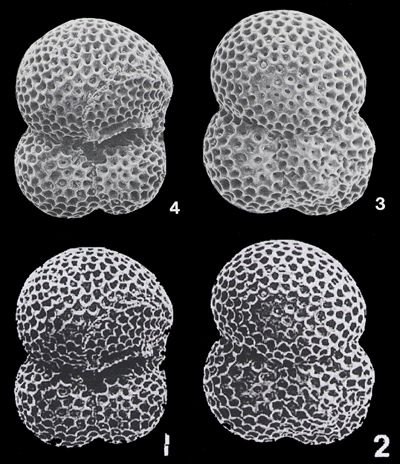
The primary additions supplementing 's work concern planktonic foraminifera. They are summarized in the following tables:
| Original photomicrographs | (2003) | |||
| (1983) | Pl. 4, fig. 4 | Subbotina triloculinoides see Fig. 6 |
Pl. 2, fig. 1 | Globigerinoides trilobus see Fig. 6 |
| Pl. 4, fig. 3 | Subbotina triloculinoides see Fig. 6 |
Pl. 2, fig. 2 | Globigerinoides immaturus see Fig. 6 |
|
| Pl. 8, fig. 11 | Globigerinoides trilobus | Pl. 2, fig. 3 | Globigerinoides trilobus | |
| Pl. 8, fig. 16 | Globigerinoides succulifer | Pl. 2, fig. 4 | Globigerinoides succulifer | |
| Pl. 8, fig. 20 | Globigerinoides ruber | Pl. 2, fig. 5 | Globigerinoides subquadratus | |
| Pl. 8, fig. 27 | Globoquadrina globosa | Pl. 2, fig. 10 | Globoquadrina dehiscens | |
| Pl. 8, fig. 6 | Globigerinoides sacculifer | Pl. 2, fig. 14 | Globigerinoides succulifer (sic) | |
| & (1993) | Pl. 9, fig. 5 | Globoquadrina baroemoenensis | Pl. 2, fig. 6 | Globigerina bulloides |
| Pl. 9, fig. 9 | Globoquadrina ? cf. G. extans | Pl. 2, fig. 8 | Globigerina angustiumbilicata | |
| et alii (1993) | Pl. 9, fig. 9 | Globigerina ciperoensis | Pl. 2, fig. 7 | Globigerina ciperoensis |
| Pl. 9, fig. 10 | Globigerina ciperoensis | Pl. 2, fig. 9 | Globigerina angulisuturalis | |
| Pl. 9, fig. 1 | Globigerina angulisuturalis | Pl. 2, fig. 11 | Globigerina angulisuturalis | |
| Pl. 7, fig. 19 | Cassigerinella chipolensis | Pl. 2, fig. 13 | Cassigerinella chiploensis (sic) | |
| & (1997) | Pl. 4, fig. 1 | Subbotina triloculinoides | Pl. 2, fig. 12 | Globigerinnella (sic) obesa |
Click on thumbnail to enlarge the image.
Figure 6: Upper row: two images from (1983, both Subbotina triloculinoides); lower row: the two photomicrographs of (2003, as n° 1 labelled Globigerinoides trilobus and n° 2 called G. immaturus). Nota bene: the form illustrated here as Globigerinoides trilobus had already appeared in (2001: Fig. 6.25) as Subbotina triloculinoides. [Some rights reserved]
Though we are quite suspicious regarding the source of the illustrations for the remaining material, particularly the benthic foraminifers, we were not able to demonstrate that these photomicrographs were "lifted" from the publications of other authors.
The fraud (see definitions in , 2001, and et alii, 2006) was exposed because the author pretended that he himself illustrated his material.
Most photographic illustrations were
"borrowed" from the publications of other authors, with or without manipulation. In papers dealing with fossil red or green algae
(see Figs. 1 ![]() - 2
- 2 ![]() ,
for instance), there are obvious evidence of fabrication: cropping
(see , 1996a, Figs. 3.1 & 3.8), grouping
(see , 1996b, Figs. 3.1.a & 3.6), masking
(see , 1996b, Figs. 3.1.b, 3.2.a-b & 3.3), etc..
On the contrary, in most papers dealing with foraminifers or charophytes, the
images were lifted without significant changes, except for a flip (mirror image)
or a rotation (a number of degrees, 90°, or upside down):
,
for instance), there are obvious evidence of fabrication: cropping
(see , 1996a, Figs. 3.1 & 3.8), grouping
(see , 1996b, Figs. 3.1.a & 3.6), masking
(see , 1996b, Figs. 3.1.b, 3.2.a-b & 3.3), etc..
On the contrary, in most papers dealing with foraminifers or charophytes, the
images were lifted without significant changes, except for a flip (mirror image)
or a rotation (a number of degrees, 90°, or upside down):
the same foraminifers appear as left coiled in the first paper
and right coiled in the next paper or vice-versa (Fig. 3 ![]() ),
possibly because (first in et alii,
1988, then in et alii,
1997) neglected to assure the use of the same side of the negative;
),
possibly because (first in et alii,
1988, then in et alii,
1997) neglected to assure the use of the same side of the negative;
some gyrogonites (
in & , 1999) appear with a odd orientation (Fig. 5 ![]() ),
that is with their base oriented upward contrary to a basic rule of charophyte iconography.
),
that is with their base oriented upward contrary to a basic rule of charophyte iconography.
Such errors demonstrate 's woeful ignorance of the conventions in both fields of micropaleontology. If the first example is not easy to detect by the reviewers, the second should have alerted any charophyte-expert, if one had been requested to review the manuscript before its publication.
In several cases, even images of type material (holotypes, paratypes, topotypes) were copied. Paleontologists know that the name of a fossil is attached to its type specimen and that this material is commonly used as the reference for comparison with new findings, therefore choosing photomicrographs of holotypes as did repeatedly is possibly the most stupid of his falsifications:
Again such plagiarism should have alerted experts on charophytes or on planktonic foraminifers, if either had been asked to evaluate these manuscripts before their publication. Duplicated photomicrographs in some publications are commonly due either to a careless mistake or to inadequate knowledge of the studied field (see for instance et alii1986): figure 1 in their Pl. 1 is the exact copy of figure 2 in the same plate, but it is rotated 90° clockwise).
Some microfossils were designated by a specific name other than that ascribed it originally (see tables above and in et alii, 2008). commonly altered valid names of species to make them conform to Cenozoic charophyte biozonation, a fact which testifies to his dishonest intent.
Certain aberrancies in stratigraphic distribution should have warned specialists about the low degree of credibility to be accorded these articles and all the works of this author. For instance, the green algae Otternstella lemmensis () (formerly known as Heteroporella) and Salpingoporella annulata had never been reported in strata younger than Valanginian; their recording by (1996b) in Upper Cretaceous strata ought to have warned the specialists about a misidentification; actually it did so but we could hardly have imagined that the specimens illustrated were not from the author's collection. Charophyte gyrogonites were reported by from stratigraphic intervals other than the interval supposedly studied. As such the misdated co-occurrence of Late Eocene and Early Miocene gyrogonites in Upper Eocene sediments ( & , 1999) and that of specimens of Mid Jurassic, Early Cretaceous and Early Miocene age in one locality (, 2000) should have puzzled the reviewers.
According to & (1999, p. 1, lines 24-25 of their Abstract) their specimens were "illustrated for the first time" from remote localities in Egypt and Libya, from which additional samples would not be easy to obtain. Because none of the figured specimens actually came from Egyptian and Libyan localities the existence of these microfossils and even of the strata supposedly sampled becomes problematic. In this regard, it is significant that no redepositories are listed for any of these specimens. Consequently, and obviously by design, verification of 's "findings" is not possible.
's fabricated data started polluting later publications and might have affected to some degree the validity of their conclusions (see for instance the recent papers of 2003), et alii2003), & 2006), et alii2005, 2006) and & 2006).
The issue of image verification should become mandatory soon, for
conventional methods of photography (emulsions on film of halides of silver) are
being replaced by electronic methods (Fig. 7 ![]() )
that are even easier to manipulate ( et alii,
2006).
)
that are even easier to manipulate ( et alii,
2006).
Click on thumbnail to enlarge the image.
Figure 7: Find of a giant planktonic foraminifer in a remote area of the Middle East. An obvious montage made with a common photo editor. The particular example is easily detectable because pixel sizes in the paste-up vary widely.
The most regrettable aspect of these frauds is that the discredit tarnishes not only the coauthors who, in other cases of this type (the frauds, brought to light by et alii, 1988, supplemented by , 1989), have been presumed innocent. Publishers should try prevent such misconduct, not only for legal reasons (infringement of the copyright rules) but also because it tarnishes the reputation of established journals and their editorial committees. This fraud had the "merit" of revealing a flaw in our system of evaluation for contributions to scientific publications. What would have occurred if the author had chosen not to illustrate "his" material? This poses the question: What degree of credibility should be given to publications without illustrations or, as in a case recently documented by et alii (2007), that came with poor illustrations? To insure against fraud editors should demand that authors document the material with photos (whether or not the manuscript is illustrated by their use) that provide the bases for publication (or at least its most significant elements) and that they indicate a public site (Museum, national collection, etc.) where this material, properly referenced, will be permanently accessible to the scientific community. However one reviewer (R.S.) reminded us that "a basis for good science is trust" (we agree); he also stated that such "drastic measures" will not be beneficial to science in introducing "more bureaucracy". While reviewing a manuscript submitted to the Revista Española de Micropaleontología (2004) was able to identify an image pirated from his own work ( et alii, 1993); thanks to this image identification it was then possible to ask the author to retract his submission. In conclusion, reviewers are -and will remain- the keystone in evaluation (see , 2001; et alii, 2006; , 2007). But, if the submittal passes peer-review undetected, subsequent exposure through later identification of pirated material remains probable ( et alii, 2006), as demonstrated herein.
Carnets' publications are usually licensed under the Creative Commons Attribution 2.5 License. But the material illustrated herein was de facto published earlier in other journals and some rights are therefore reserved. Special thanks are due to the editors of Géologie méditerranéenne, Journal of African Earth Sciences, Journal of foraminiferal Research, Revue de Paléobiologie, ... who granted us permission to re-use this material. Alternatively the "fair use" clause fully applies as it was implemented to better document the fraud. The first author (B.G.) would like to thank the many persons who unreservedly supported him in his quest to unearth this blatant and long-lived fraud. Thanks are due too to Michel and Robert for their accurate quality check of the manuscript, and special thanks to Nestor who helped make this critique easier to read.
Remark: Starred references (*) were listed in the bibliographic references of 's papers.
P.A. (2001).- Academic misconduct, definitions, legal issues, and management. In: A. & M.M. (Eds.), Expanding Horizons in Teaching and Learning.- Proc. 10th Annual Teaching Learning Forum, Perth, 2001 (Curtin University of Technology)
URL: http://lsn.curtin.edu.au/tlf/tlf2001/addison2.html
J. (2004).- Plagiarism in palaeontology. A new threat within the scientific community.- Revista Española de Micropaleontología, Madrid, vol. 36, n° 2, p. 349-352.
* J., J.C. & J.M. (1993).- Algal nodules in the Upper Pliocene deposits at the coast of Cadiz (S Spain).- In: F., P. & M. (eds.), Studies on fossil benthic algae.- Bollettino della Societá Paleontologica Italiana, Modena, Special Volume, n° 1, p. 1-7.
* W.A. & R.N. (1997).- Biostratigraphy, phylogeny and systematics of Paleocene trochospiral planktic foraminifera.- Micropaleontology, New York, vol. 43, supplement 1, 116 p.
M., L., J., B. & R. (2007).- Comment to "Latest-Cretaceous/Paleocene karsts with marine infillings from Languedoc (South of France); paleogeographic, hydrogeologic and geodynamic implications by P.J. et al.".- Geodinamica Acta, Paris, vol. 20, n° 1, p. 403-413.
X. (2004a).- Fallout from fraud.- The Scientist, Philadelphia, vol. 5, n° 1, 20040922-02
URL: http://www.the-scientist.com/news/20040922/02/
X. (2004b).- Plagiarism in paleontology.- The Scientist, Philadelphia, vol. 5, n° 1, 20041008-03
URL: http://www.the-scientist.com/news/20041008/03/
N., M.A. & R. (2006).- Salpingoporella, a common genus of Mesozoic Dasycladales (calcareous green algae).- Revue de Paléobiologie, Genève, vol. 25, n° 2, p. 457-517.
W.P. & R.M. (1993).- 10. High-resolution Neogene planktonic foraminifer biostratigraphy of Site 806, Ontong Java Plateau (western Equatorial Pacific).- Proceedings of the Ocean Drilling Program, Scientific Results, College Station, vol. 130, p. 137-178.
P.G., H.A.B. & S. (2004).- Editorial.- Journal of African Earth Sciences, Oxford, vol. 40, n° 3-4, p. 113-114.
M. & N. (1984).- New Porocharaceae from the Bathonian of Europe: phylogeny and palaeoecology.- Palaeontology, London, vol. 27, part 2, p. 295-305.
M. & M. (1977).- Étude biostratigraphique et paléobotanique (Charophytes) des formations continentales d'Aquitaine, de l'Éocène supérieur au Miocène inférieur.- Bulletin de la Société géologique de France, Paris, (7e série), t. XIX, n° 2, p. 341-354, pls. X-XIII.
M. (1977).- Étude floristique et biostratigraphique des Charophytes dans les séries du Paléogène de Provence.- Géologie méditerranéenne, Marseille, t. IV, n° 2, p. 109-138.
E. (1979).- Paleoecology and microfacies of Permian, Triassic and Jurassic algal communities of platform and reef carbonates from the Alps.- Bulletin des Centres de Recherches Exploration-Production Elf-Aquitaine, Pau, vol. 3, n° 2, p. 529-587.
* E. (1982).- Microfacies analysis of limestones.- Springer-Verlag, New York, 633 p.
M. (2003).- Miocene corals from Wadi El Hommor, Sinai (Egypt).- Neues Jahrbuch für Geologie und Paläontologie, Abhandlungen, Stuttgart, Band 229, p. 159-187.
N. (1980).- Les Charophytes du Montien de Mons (Belgique).- Review of Palaeobotany and Palynology, Amsterdam, vol. 30, p. 67-88.
L. & N. (1981).- Étude sur les Charophytes tertiaires d'Europe Occidentale. III - Le genre Gyrogona.- Paléobiologie continentale, Montpellier, vol. XII, n° 2, 35 p. + pls. I-VI.
B. (2007).- Impact of research assessment on scientific publication in Earth Sciences. In: Assessing the quality and impact of research: practices and initiatives in scholarly information.- ICSTI - The International Council for Scientific and Technical Information, 2007 Public Conference, Nancy, France, June 22th-20th, 2007.
URL: http://international.inist.fr/article183.html#granier
B., M., I.I. & B. (2007).- Further investigation of the 's micropaleontological fraud: The stolen algae. In: T. & I. (eds.), Field Trip Guidebook and Abstracts.- 9th International Symposium on Fossil Algae - Croatia 2007, Zagreb (September 19th-20th), Hrvatski geološki institut - Croatian Geological Survey, p. 223-224.
B., M., P.G. & S.M. (2008, available online 7 October 2007).- A micropalaeontological fraud that affected the JAES.- Journal of African Earth Sciences, Oxford, vol. 50, n° 1, p. 1-5.
M.M. (1996a).- Coralline red algae from the Middle Miocene deposits of Gebel Gushia west-central Sinai, Egypt.- Neues Jahrbuch für Geologie und Paläontologie, Abhandlungen, Stuttgart, Band 199, p. 1-15.
M.M. (1996b).- Dasycladacean algae from the Turonian Wata Formation, Gebel Um Heriba, West-Central Sinai, Egypt.- Neues Jahrbuch für Geologie und Paläontologie, Monatshefte, Stuttgart, Heft 9, p. 527-538.
M.M. (1998).- Clavatorella salumensis, a new species of planktonic foraminiferal from the Early Pliocene of the Wadi El Salum area, northern Western Desert, Egypt.- Neues Jahrbuch für Geologie und Paläontologie, Monatshefte, Stuttgart, Heft 10, p. 595-603.
M.M. (1999).- Lithostratigraphy and planktonic foraminiferal biostratigraphy of the Late Eocene-Middle Miocene sequence in the area between Wadi Al Zeitun and Wadi Al Rahib, Al Bardia area, northeast Libya.- Journal of African Earth Sciences, Oxford, vol. 28, n° 3, p. 619-639.
M.M. (2000).- On the occurence and significance of Charophyte from Ar Rajban Member, Kiklah Formation, Wadi Ar Rajban, Mizdah Area, Northwest of Libya.- Arab Gulf Journal of Scientific Research, Al Manamah, vol. 18, n° 3, p. 173-183.
M.M. (2001).- Biostratigraphy of the Upper Cretaceous Lower Eocene succession in the Bani Walid area, northwest Libya.- Journal of African Earth Sciences, Oxford, vol. 33, n° 1, p. 69-89.
M.M. (2002).- Biostratigraphy and paleoecology of the Early Pliocene sequence, Salum area, north Western Desert, Egypt.- Revista Española de Micropaleontología, Madrid, vol. 34, n° 1, p. 19-38.
M.M. (2003).- Contribution to the stratigraphy and paleontology of the Miocene sequence at Al Khums area, Sirte basin, NW Libya.- Revista Española de Micropaleontología, Madrid, vol. 35, n° 2, p. 195-228.
M.M. & M.A. (2000).- Stratigraphy and microfacies of the Oligocene sequence at Gabal Bu Husah, Marada Oasis, south Sirte Basin, Libya.- Facies, Erlangen, 42, p. 93-106.
M.M. & A.A. (2000).- Stratigraphy and facies analysis of the Miocene sequence at Gabal Hammam Sayidna Musa and Wadi Abura, southern Sinai, Egypt.- Neues Jahrbuch für Geologie und Paläontologie, Monatshefte, Stuttgart, Heft 7, p. 385-408.
C.A.L., R.L., I.D. & I.R. (2005).- Normal faulting as a control on the stratigraphic development of shallow marine syn-rift sequences.- Sedimentology, Oxford, vol. 52, n° 2, p. 313-338.
C.A.L., R.L., C.W. & I.R. (2006).- Rift-initiation development of normal fault blocks: insights from the Hammam Faraun fault block.- Journal of the Geological Society, London, vol. 163, n° 1, p. 165-183.
* J.H. (1954).- Fossil calcareous algae from Bikini atoll.- Geological Survey Professional Papers, Washington, 260M, p. 537-545.
* J.H. (1957).- Geology of Saipan, Mariana Islands. Calcareous algae.- Geological Survey Professional Papers, Washington, 280E, p. 209-246.
* J.H. (1961).- Limestone-building algae and algal limestones.- Colorado School of Mines, Boulder, 297 p.
J.H. (1963).- The algal genus Archaeolithothamnium and its fossil representatives.- Journal of Paleontology, Tulsa, vol. 37, n° 1, p. 175-211.
J.H. (1971).- An introduction to the study of organic limestones.- Quaterly of the Colorado School of Mines, Boulder, vol. 66, n° 2, 185 p.
J.H. & B.J. (1949).- Tertiary coralline algae from the Dutch East Indies.- Journal of Paleontology, Tulsa, vol. 23, n° 2, p. 193-198.
J.H. & B.J. (1950).- Tertiary and Pleistocene coralline algae from Lau, Fiji.- Bernice P. Bishop Museum (Honolulu), Bulletin, n° 201, 27 p.
J.P. & M.S. (1983).- Neogene planktonic foraminifera. A phylogenetic atlas.- Hutchinson Ross Publishing Company, Stroudsburg, 265 p.
H., H.A. & E. (1986).- Fossil algae and biostratigraphy of the Middle Eocene rock succession at the southeast of Minia, Nile Valley, Upper Egypt.- Arab Gulf Journal of Scientific Research, Al Manamah, vol. 4, n° 1, p. 141-157.
W., E. & J. (2003).- Patterns of Phanerozoic carbonate platform sedimentation. Carbonate platforms changed substantially in spatial extent, geometry, composition and palaeogeographical distribution through the Phanerozoic.- Lethaia, Oslo, vol. 36, n° 3, p. 195-225.
* R.M., C. & M.G. (1993).- 9. Oligocene planktonic foraminifer biostratigraphy of Hole 803D (Ontong Java Plateau) and Hole 628A (Little Bahama Bank), and comparison with the southern high latitudes.- Proceedings of the Ocean Drilling Program, Scientific Results, College Station, vol. 130, p. 113-136.
C.W. & R.L. (2006).- Sedimentology of rift climax deep water systems; Lower Rudeis Formation, Hammam Faraun Fault Block, Suez Rift, Egypt.- Sedimentary Geology, Amsterdam, vol. 191, n° 1-2, p. 67-87.
* C. & N. (1986).- Les Charophytes du Crétacé inférieur de la région du Maestrat (Chaîne Ibérique - Catalanides, Espagne).- Paléobiologie continentale, Montpellier, vol. XV, 66 p. + pls. I-X.
* S.M. (1991).- Dasycladacean algae from the Jurassic and Cretaceous of central Saudi Arabia.- Micropaleontology, New York, vol. 37, n° 2, p. 183-190.
* S.M. (1992).- Further record of Jurassic, and Cretaceous fossil algae from central Saudi Arabia.- Revue de Paléobiologie, Genève, vol. 11, n° 2, p. 373-383.
S.W. (1983).- Gulf of Guinea planktonic foraminiferal biochronology and geological history of the South Atlantic.- Journal of foraminiferal Research, Washington, vol. 13, n° 1, p. 32-59.
J. (1926).- Les Mélobésiées dans les calcaires crétacés de la Basse-Provence.- Mémoires de la Société géologique de France, Paris, (Nouvelle Série), t. III, fasc. 2, n° 6, 32 p., pls. I-X.
* G., M.M. & G.I. (1997).- Planktonic foraminiferal biostratigraphy of the Miocene sequence in the area between Wadi El-Tayiba and Wadi Sidri, west central Sinai, Egypt.- Journal of African Earth Sciences, Oxford, vol. 25, n° 3, p. 435-451.
R. (2006).- Trinocladus divnae and Montiella filipovici - a new species (Dasycladales, algae) from the Upper Cretaceous of the Mountain Paštrik (Mirdita Zone).- Geološki anali Balkanskog poluostrva, Beograd, Knjiga LXVII, p. 65-87.
A.A. & M.M. (1999).- The Tayiba Red Beds: transitional marine-continental deposits in the precursor Suez Rift, Sinai, Egypt.- Journal of African Earth Sciences, Oxford, vol. 28, n° 3, p. 487-506.
D. & the Editorial Policy Committee (2006).- CSE's White Paper on Promoting Integrity in Scientific Journal Publications.- Council of Science Editors, Reston, iv + 71 p.
URL: http://www.councilscienceeditors.org/editorial_policies/white_paper.cfm
J.B. & F.M. (2006).- An abelisaurid (Dinosauria: Theropoda) tooth from the Lower Cretaceous Chicla formation of Libya.- Journal of African Earth Sciences, Oxford, vol. 46, n° 3, p. 240-244.
* I. (1994).- The paleoecological implications of the charophyte flora of the Trinity Division, Junction, Texas.- Journal of Paleontology, Lawrence, vol. 68, n° 5, p. 1145-1157.
* M.S. & J.P. (1974).- A planktonic foraminifer (Clavatorella) from the Pliocene.- Journal of Foraminiferal Research, Lawrence, vol. 4, n° 2, p. 77-79.
* M.S. & J.P. (1975).- The status of Bolliella, Beella, Protentella and related planktonic foraminifera based on the ultrastructure.- Journal of Foraminiferal Research, Lawrence, vol. 5, n° 3, p. 155-165.
T. & D. (1980).- Faziesmuster oberjurassischer Plattform-Karbonate (Plassen-Kalke, Nördliche Kalkalpen, Steirisches Salzkammergut, Österreich).- Facies, Erlangen, 2, p. 241-284.
J.A. (1989).- The case of the peripatetic fossils.- Nature, London, vol. 338, n° 6217, p. 613-615.
J.A., R.K., A.K & J.W. (1988).- Silurian and Devonian of India, Nepal and Bhutan: biostratigraphic and palaeobiogeographic anomalies.- Courier Forschungsinstitut Senckenberg, Frankfurt am Main, 106, 57 p.
V.J. & S.W. (1986).- Planktonic foraminiferal biostratigraphy of the Pungo River Formation, southern Onslow Bay, North Carolina continental shelf.- Journal of foraminiferal Research, Washington, vol. 16, n° 1, p. 9-23
* E.A.A., S.E. & M. (1988).- Stratigraphy and microfacies of the Miocene sequence at Gebel Sarbut El Gamal, west-central Sinai, Egypt.- Neues Jahrbuch für Geologie und Paläontologie, Abhandlungen, Stuttgart, Band 177, p. 225-242.