◄ Carnets Geol. 22 (6) ►
![]()
Outline:
[1. Introduction and setting]
[2. Material and methods]
[3. Geology]
[4. Description]
[5. Concluding remarks]
and ...
[Bibliographic references]
Department of Paleontology, Calvert Marine Museum, P.O. Box 97, Solomons, MD 20688 (U.S.A.);
Department of Paleobiology, National Museum of Natural History, Smithsonian Institution, Washington, DC, 20013-7012 (U.S.A.)
Department of Paleobiology, National Museum of Natural History, Smithsonian Institution, Washington, DC, 20013-7012 (U.S.A.)
Department of Vertebrate Paleontology, American Museum of Natural History, 200 Central Park West, New York, NY 10024 (U.S.A.)
Published online in final form (pdf) on May 10, 2022
DOI 10.2110/carnets.2022.2206
![]()
[Editor: Alberto Collareta; technical editor: Bruno R.C. Granier]
![]()
An isolated and deformed lower tooth plate of an eagle ray (Aetomylaeus sp., Myliobatidae, Myliobatiformes) is reported herein from the Miocene deposits along Calvert Cliffs, Maryland, U.S.A. Deformed myliobatid tooth plates like this, either fossil or modern, are exceedingly rare. All medial teeth are deformed/skewed such that the right side of each tooth forms an angle of about 10 degrees to the transverse axis of the dental plate. The skewed abnormal form of the teeth in USNM PAL 726325 is not thought to be the result of modern or taphonomic deformation. Rather, the deformity exhibited by USNM PAL 726325 gives every indication that successive similarly deformed teeth came about as a result of a persistent anatomical deformity of the dental lamina. From the consistency in the deformed shape of each tooth, it would appear as though the dental lamina retained this anomalous shape at least throughout the time represented by the age of the tooth plate. If this interpretation is correct, the deformity was not fatal and did not significantly impair the functionality of the tooth pavement over a protracted part (or all) of the individual's life span.
• deformed dental lamina;
• eagle ray;
• Aetomylaeus;
• Myliobatidae;
• Miocene;
• Calvert Cliffs
Godfrey S.J., Bohaska D.J. & Maisey J. (2022).- The report of a rare deformed eagle ray (Myliobatiformes: Myliobatidae) tooth plate from the Neogene of Calvert Cliffs, Maryland, U.S.A.- Carnets Geol., Madrid, vol. 22, no. 6, p. 161-169.
Description d'un rare palet dentaire déformé de raie aigle de mer (Myliobatiformes : Myliobatidae) du Néogčne de Calvert Cliffs, Maryland, États-Unis d'Amérique.- Un palet dentaire inférieur, isolé et déformé, d'une raie aigle de mer (Aetomylaeus sp., Myliobatidae, Myliobatiformes) décrit ici provient d'un affleurement miocčne des Calvert Cliffs, Maryland, États-Unis d'Amérique. Les palets dentaires déformés de myliobatides tels que celui-ci, qu'ils soient fossiles ou actuels, sont extręmement rares. Toutes les dents mésiales sont déformées ou asymétriques, de telle maničre que le côté droit de chaque dent forme un angle d'environ 10 degrés sur l'axe transverse du palet dentaire. La forme anormale asymétrique de la dent USNM PAL 726325 n'est pas considérée comme résultant d'une déformation taphonomique ou actuelle. Mais la déformation présentée par USNM PAL 726325 indiquerait plutôt que des dents successives, pareillement déformées, surviennent comme le résultat d'une déformation anatomique récidivante du palet dentaire. D'aprčs la constance de la silhouette déformée de chaque dent, il apparaîtrait que le palet dentaire a conservé sa forme anormale au moins au cours de la période représentée par l'âge de la plaque dentaire. Si cette interprétation est juste, cette difformité n'était pas fatale et n'handicapait pas de maničre significative la fonctionnalité du pavage dentaire pendant une partie notable de (voire toute) l'espérance de vie de l'individu.
• palet dentaire déformé ;
• raie aigle de mer ;
• Aetomylaeus ;
• Myliobatidae ;
• Miocčne ;
• Calvert Cliffs
In the case study presented here, we describe a Miocene epoch lower tooth plate of the extant myliobatid genus, Aetomylaeus sp. Garman, 1908, that exhibits a right-side anteriorly-skewed medial tooth deformity. Anomalous dentitions like this are exceedingly rare in rays in general and hitherto have only been described in three specimens figured by Hovestadt and Hovestadt-Euler (2013, see below). These latter specimens are not deformed to the same degree as the one featured herein. Our aim is not merely to document such an occurrence in this taxon, but also to highlight the extent to which such a deformity can affect replacement tooth formation.
Eagle rays are large
aquilopelagic (forms with wing-like discs) chondrichthyans that are powerful
swimmers and durophagous predators consuming gastropods and bivalves, as well as
decapod crustaceans (Last et al., 2016). They have rigid jaws and large muscles that generate bite forces capable
of fracturing mollusk shells (Kolmann et al., 2015; Villafańa et
al., 2019). Eagle rays of the genus Aetomylaeus are known from
the late Eocene to the present (Villafańa et al.,
2019). However, they are most common in the Neogene (Carrillo-Briceńo
et al., 2019; Villafańa et
al., 2019). Villafańa et al.
(2019) provide a detailed listing of where geographically, and when geologically
specimens attributed to Aetomylaeus
have been found. Aetomylaeus is the
sole myliobatid known from the fossil-rich Calvert Cliffs (Miocene, Chesapeake
Group) located along the western shore of Chesapeake Bay (Fig. 1 ![]() ) in Calvert
County, Maryland, U.S.A. (Kent, 2018) [Note that Kent (2018)
mentioned two myliobatid genera, i.e.,
Pteromylaeus and Aetobatus. Now however, Pteromylaeus =
Aetomylaeus, and Aetobatus has been transferred to the Aetobatidae].
) in Calvert
County, Maryland, U.S.A. (Kent, 2018) [Note that Kent (2018)
mentioned two myliobatid genera, i.e.,
Pteromylaeus and Aetobatus. Now however, Pteromylaeus =
Aetomylaeus, and Aetobatus has been transferred to the Aetobatidae].
|
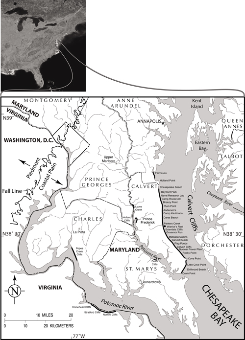
Figure 1:
Satellite image (insert) of Eastern North America and below a portion of the
northern section of Chesapeake Bay and parts of the States of Maryland and
Virginia. The
one black dot (immediately south of Parkers Creek, Calvert County, MD) marks the
location where the Miocene stingray pathological mouthplate was found. Modified from Vogt et al.
(2018). |
The
dentition of Aetomylaeus spp. consists
of flat-crowned teeth that
interlock to form pavement-like tooth plates (Cappetta, 2012, Fig. 441 F; Hovestadt
and Hovestadt-Euler, 2013; Kolmann et al.,
2015, Fig. 1; Kent, 2018, Fig. 2.25; Marramŕ et al.,
2019, Fig. S4b). Each tooth forms from a
continuous dental lamina, with new teeth
developing through a conveyor belt-like replacement mechanism (Underwood et
al., 2015; Rasch et al.,
2020). In Aetomylaeus,
individual upper and lower teeth are not distinct morphologically (Cappetta,
2012; Kent, 2018, Fig. 2.25; Marramŕ et al.,
2019, Fig. S4b). However, upper and lower tooth pavements
can be distinguished on the basis of their anteroposterior curvature (Hovestadt
and Hovestadt-Euler, 2013, Pl.
14; Kolmann et al., 2015, Fig. 1, sagittal view). Lower tooth pavements are
nearly flat to only gently convex dorsally (like the one described herein), whereas upper
dentitions are much more strongly convex dorsally from front to back (Kolmann
et
al., 2015, Fig. 1, sagittal view; Villafańa et
al., 2019, Fig. 2a). Only the anterior parts of the upper and lower tooth
plates occlude to form a functional grinding pad, as evidenced by the wear
pattern visible on the tooth plate (Figs. 2 ![]() - 3
- 3 ![]() ).
).
|
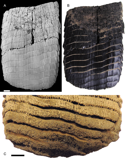
Figure 2:
USNM PAL 726325 eagle ray (Aetomylaeus
sp.) partial tooth plate. A and B are both occlusal views. A. Specimen
whitened with sublimed ammonium chloride to highlight surface detail. C.
Posterior margin of the tooth plate in a basal view. Scale bars equal 10 mm. |
|
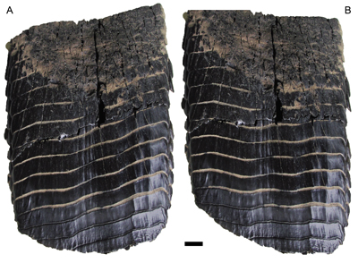
Figure 3:
USNM PAL 726325 eagle ray (Aetomylaeus
sp.) partial tooth plate seen in occlusal view. A. Tooth plate showing its
natural configuration preserving the semi-circular wear pattern on its anterior
margin. B. Tooth plate altered in Photoshop® to correct for the right-hand skew.
Now the wear pattern is skewed towards the posterior right. Scale bar equals 10
mm. |
USNM PAL 726325, Department of Paleobiology collections, National Museum of Natural History, Smithsonian Institution, Washington, DC, U.S.A. Tooth terminology follows Cappetta (2012).
There
was no sediment adhering to the three pieces of the mouth plate when they were
found. Polyvinyl butyral in acetone was used to glue the three pieces together,
but otherwise, no consolidant was applied to the specimen. In its natural state,
USNM PAL 726325 is nearly jet
black (Fig. 2.B ![]() ), so to improve
contrast and visibility (Fig. 2.A
), so to improve
contrast and visibility (Fig. 2.A ![]() ), the
mouth plate was lightly dusted with
sublimed ammonium chloride (a whitening technique described by Cooper
(1935) and Feldman (1989)). After the specimen was photographed with a
Nikon Coolpix P510 camera on black velvet under fluorescent light, the ammonium
chloride was removed by holding the specimen under running water (Shelburne
& Thompson, 2016). As a cautionary note, specimens coated with ammonium
chloride should be stable enough to withstand a fresh water rinse because
without that, there would be the possibility of a residue of hydrochloric acid (HCl)
left on the specimens if they are not washed thoroughly.
), the
mouth plate was lightly dusted with
sublimed ammonium chloride (a whitening technique described by Cooper
(1935) and Feldman (1989)). After the specimen was photographed with a
Nikon Coolpix P510 camera on black velvet under fluorescent light, the ammonium
chloride was removed by holding the specimen under running water (Shelburne
& Thompson, 2016). As a cautionary note, specimens coated with ammonium
chloride should be stable enough to withstand a fresh water rinse because
without that, there would be the possibility of a residue of hydrochloric acid (HCl)
left on the specimens if they are not washed thoroughly.
The images were edited in Adobe Photoshop® and compiled in Adobe Illustrator®.
USNM
PAL 726325 was not found in
situ. Rather, it was found in three pieces on the beach over a two-month
period. The first piece was found by
author D.J.B. on 14 January 2018, 85 m north of the "waterfall", high on the
beach. In a form going back to at least Remington Kellogg, this is 85 m north of
the south end of the third cliff south of the mouth of Parkers Creek, Port
Republic, Calvert County, Maryland (Fig. 1 ![]() ). Approximate GPS coordinates are:
N
38°31.797' W 076°30.970'. In an occlusal view of the dental plate, this
piece comprises the upper left quarter. The second piece was found on 20 January
2018, 17 m north of the "waterfall", in the water, in gravel, at the
water's edge. Approximate GPS coordinates are: N 38°31.767', W 076°30.955'.
Therefore, the two pieces were found approximately 72 m apart. This is the
largest piece comprising the lower half of the plate. The third piece (upper
right) was apparently found in the same area by Joseph Cumberland, 11 February
2018.
). Approximate GPS coordinates are:
N
38°31.797' W 076°30.970'. In an occlusal view of the dental plate, this
piece comprises the upper left quarter. The second piece was found on 20 January
2018, 17 m north of the "waterfall", in the water, in gravel, at the
water's edge. Approximate GPS coordinates are: N 38°31.767', W 076°30.955'.
Therefore, the two pieces were found approximately 72 m apart. This is the
largest piece comprising the lower half of the plate. The third piece (upper
right) was apparently found in the same area by Joseph Cumberland, 11 February
2018.
These pieces of the dental plate were beach-float long enough for any adhering matrix to wash off. Determination of source bed from lithology or microfossils is therefore not possible. Given their sizes and good preservational condition, the pieces probably did not travel far from their originating position in the cliffs. In the immediate area where USNM PAL 726325 was found, only Shattuck-Zones 11-19 are exposed in the cliffs, which includes the upper portion of the Plum Point Member (Shattuck-Zones 11-15) of the Calvert Formation along with the Drumcliff (Shattuck-Zone 17), St. Leonard (Shattuck-Zone 18), and Boston Cliff (Shattuck-Zone 19) members of the Choptank Formation (Kidwell et al., 2015, Fig. 15). Kent (2018) recently compiled a detailed review of the diverse chondrichthyan fauna known from Calvert Cliffs. Aetomylaeus (= Pteromylaeus) has been reported (Kent, 2018, p. 128) in situ in various (but not all) Shattuck-Zones 1 through 23, including 11-12, 14, 17, 19, which cover the span of Shattuck-Zones in the adjacent cliff section, as well as sections north and south. We therefore conclude that USNM PAL 726325 originated from within Shattuck-Zones 11 through 19, but cannot restrict it further. The sediments comprising this section of the cliffs range in age between approximately 16.5-12.5 Ma (Perez et al., 2019, Fig. 1). Kidwell et al. (2015, and references therein) provide a detailed description of the geology of the cliffs and Vogt et al. (2018) give an overview of the very diverse Miocene (mostly marine) biota that is preserved along Calvert Cliffs.
USNM PAL 726325 consists
of a lower dental plate that has a maximum midline length and width of 135 mm
and 101 mm respectively. Some small fragments are missing where the plate broke
apart. There is a shallow longitudinal concavity medially that extends the full
length of the tooth plate, as opposed to the convex surface expected. The dental
plate consists mostly of 15 elongate medial teeth (= symphyseal teeth; Underwood
et al., 2015), the posterior three of
which are not complete width-wise (Figs. 2 ![]() - 3
- 3 ![]() ). It is not known how many more
medial teeth may have been present in life. A smaller number of lateral teeth
are preserved. They are much shorter width-wise and lozenge-shaped, but
otherwise have a structure comparable to that of the medial teeth (Hovestadt
& Hovestadt-Euler, 2013; Kent, 2018; Villafańa et
al., 2019).
Although many lateral teeth are missing, at two positions on the left and one
right, two files of teeth are present. There were therefore at least five tooth
files in this individual.
). It is not known how many more
medial teeth may have been present in life. A smaller number of lateral teeth
are preserved. They are much shorter width-wise and lozenge-shaped, but
otherwise have a structure comparable to that of the medial teeth (Hovestadt
& Hovestadt-Euler, 2013; Kent, 2018; Villafańa et
al., 2019).
Although many lateral teeth are missing, at two positions on the left and one
right, two files of teeth are present. There were therefore at least five tooth
files in this individual.
The medial teeth are over nine times wider than long (a characteristic of the genus Aetomylaeus), whereas teeth of Myliobatis are only four to five times wider than long (Kent, 2018). The maximum width of the anterior-most medial tooth (as measured along the width of the skewed tooth) is 98.5 mm, whereas the same dimension of the posterior-most complete tooth is 94.5 mm. Therefore, the medial teeth decrease in width by 4 mm from front to back.
The
surface of the medial tooth crowns is lightly ornamented, mostly with
labiolingually oriented low ridges, more numerous and conspicuously developed
laterally (Fig. 2.A-B ![]() ). The roots of fully formed teeth are polyaulacorhizous,
with 65 fine, labiolingually directed laminae separating the nutritive groves (Fig.
2.C
). The roots of fully formed teeth are polyaulacorhizous,
with 65 fine, labiolingually directed laminae separating the nutritive groves (Fig.
2.C ![]() ). The occlusal surface at the anterior end of the dental plate
preserves a semi-circular wear pattern (Figs. 2.A-B
). The occlusal surface at the anterior end of the dental plate
preserves a semi-circular wear pattern (Figs. 2.A-B ![]() ,
3
,
3 ![]() ) that presumably developed
in life where opposing dentitions crushed prey.
) that presumably developed
in life where opposing dentitions crushed prey.
In an occlusal view of a normal tooth plate (Kent, 2018, Fig. 2.25), a
line passing through the maximum width of each medial tooth would be orthogonal
to the long axis of the plate. However, in USNM PAL 726325, adjacent medial teeth are skewed such that the right side
of each tooth forms an angle of about 10 degrees to the transverse axis of the
dental plate (Figs. 2 ![]() - 3
- 3 ![]() ).
).
In a basal view of the posterior margin of the dental plate
(Fig. 2.C ![]() ),
parts of four incomplete (widthwise) teeth are preserved. Although the occlusal
surface of these teeth gives every appearance of having been fully mineralized
in life (Fig. 2.A-B
),
parts of four incomplete (widthwise) teeth are preserved. Although the occlusal
surface of these teeth gives every appearance of having been fully mineralized
in life (Fig. 2.A-B ![]() ), the expected polyaulacorhizous
basal surface of each tooth appears to have been eroded and is poorly defined.
Perhaps the laminae of these young teeth had not yet fully mineralized at the
time of death, i.e., they consisted of
soft tissue, which did not preserve (favored interpretation), or they were
eroded as a consequence of a taphonomic process, (perhaps, although this would
appear to be less likely given that the polyaulacorhizous
roots further forward on the tooth plate are fully formed).
), the expected polyaulacorhizous
basal surface of each tooth appears to have been eroded and is poorly defined.
Perhaps the laminae of these young teeth had not yet fully mineralized at the
time of death, i.e., they consisted of
soft tissue, which did not preserve (favored interpretation), or they were
eroded as a consequence of a taphonomic process, (perhaps, although this would
appear to be less likely given that the polyaulacorhizous
roots further forward on the tooth plate are fully formed).
Chondrichthyan tooth abnormalities were first described in the late 1700s. Andre (1784) figured a jaw of extant Galeocerdo that had been pierced by a stingray tail spine. Delfortrie (1877) and Woodward (1888) also noted dental anomalies in fossil forms. Much more recently, Cadenat (1962) described and figured eight kinds of feeding-related dental injuries/abnormalities (Types 1-8) in extant chondrichthyans. Becker et al. (2000) surveyed 200 modern lamniform and carcharhiniform sharks, concluding that chondrichthyan tooth deformities come about as a result of feeding-related injury to the dental lamina, particularly by impaction of chondrichthyan and teleost fin and tail spines. Likewise, Balbino and Antunes (2007) described chondrichthyan tooth deformities that they thought were caused by damage to the dental lamina by hard foreign objects, like bone or ray spines. Cappetta (2012, Fig. 20) also described and figured a variety of chondrichthyan tooth abnormalities, but none in rays. Gudger (1933) described unusual dentitions in rays. In the Myliobatidae, the dental abnormalities that he described consisted almost entirely of asymmetrical differences in tooth counts, only within the genus Myliobatis. Three published specimens show some similarity to USNM PAL 726325. They include a specimen attributed to the extant Aetobatus narinari (Hovestadt & Hovestadt-Euler, 2013, Pl. 14, fig. 1a), the fossil "Aetobatis" irregularis (Agassiz, 1843) (Hovestadt & Hovestadt-Euler, 2013, Pl. 28, fig. 1), where the teeth are not symmetrical, and the fossil form Myliobatis irregularis Dixon, 1850 (Hovestadt & Hovestadt-Euler, 2013, Pl. 31, fig. 1). Hovestadt and Hovestadt-Euler (2013) reported that there is strong ontogenetic heterodonty in tooth morphology within Aetomylaeus. However, the skewed abnormal form of the teeth in USNM PAL 726325 is not the result of ontogenetic or sexual heterodonty, nor is it thought to be the result of the localized impaction of fin or tail spines, nor from modern or taphonomic deformation. It would be difficult to account for such a uniform taphonomic deformity given that the teeth are for the most part intact, showing no signs of breakage. We do not know how or when in the life of this eagle ray the dental lamina became deformed. Was it an embryonic developmental irregularity/perturbation (i.e., congenital), or the result of physical trauma?
Gudger (1937), Underwood et al. (2015), and Rasch et al. (2020), all report that in sharks, skates, and rays, tooth replacement begins from the successional lamina, which is continuous with the dental lamina (Gudger, 1937; Underwood et al., 2015; Rasch et al., 2020). The deformity in USNM PAL 726325 gives every indication that successive similarly deformed teeth came about as a result of a persistent anatomical deformity of the dental lamina. It is not known what caused the original deformity (e.g., if the jaw cartilage beneath the dental lamina was deformed), but evidently it interfered with tooth formation from the earliest stage in growth of the tooth germ. We know of no report of an impacted hard object resulting in such a widespread deformity. From the consistency in the deformed shape of each tooth, it would appear as though the dental lamina retained this anomalous shape, at the very least throughout the time represented by the age of the tooth plate. If this interpretation is correct, the deformity was not fatal and did not significantly impair the functionality of the tooth pavement over a protracted part (or all) of the individual's lifespan. Apparently, there are no quantitative data on the rates of tooth replacement in rays (Berkovitz & Shellis, 2017).
Although
the anterior end of the tooth plate in USNM PAL 726325
is asymmetrical, the posterior end is very nearly symmetrical (Fig.
2 ![]() ).
On a normal plate, growth is like an assembly line; one tooth or row forms and
moves forward so that the next tooth (or row) can form. Presumably, in a normal
dentition, the full width of each tooth would express/present very nearly the
same level of mineralization. However, in USNM PAL 726325 (Fig.
2.C
).
On a normal plate, growth is like an assembly line; one tooth or row forms and
moves forward so that the next tooth (or row) can form. Presumably, in a normal
dentition, the full width of each tooth would express/present very nearly the
same level of mineralization. However, in USNM PAL 726325 (Fig.
2.C ![]() ), the roots of the teeth are more fully mineralized/developed
on the right-hand side of the dentition (i.e.,
left-hand side of the image). In fact, in Figure 2.C
), the roots of the teeth are more fully mineralized/developed
on the right-hand side of the dentition (i.e.,
left-hand side of the image). In fact, in Figure 2.C ![]() , the polyaulacorhizous roots are present on the
right-hand side of the fourth tooth, whereas they are not formed in the center
or left-hand side of that tooth. Therefore, that part of the tooth that is
further forward on the conveyer belt is more completely developed, whereas the
same tooth on the left appears to be in an earlier stage of formation. Therefore,
not only does the tooth plate function properly despite the abnormality, but
also it develops symmetrically. One developmental program is turning out
individual teeth of the proper size (length and width), although at an abnormal
angle, yet the stage of development overlying it (place on the conveyer belt),
working at the same time, is seemingly ignoring the tooth boundaries. What is
remarkable about this deformity is that the teeth, although misshapen, appear to
have been fully functional in their collective ability to crush hard prey. It is
not known if the teeth in the opposing jaw were similarly deformed, but the wear
pattern at the anterior end of the tooth plate appears to be bilaterally
symmetrical (Figs. 2
, the polyaulacorhizous roots are present on the
right-hand side of the fourth tooth, whereas they are not formed in the center
or left-hand side of that tooth. Therefore, that part of the tooth that is
further forward on the conveyer belt is more completely developed, whereas the
same tooth on the left appears to be in an earlier stage of formation. Therefore,
not only does the tooth plate function properly despite the abnormality, but
also it develops symmetrically. One developmental program is turning out
individual teeth of the proper size (length and width), although at an abnormal
angle, yet the stage of development overlying it (place on the conveyer belt),
working at the same time, is seemingly ignoring the tooth boundaries. What is
remarkable about this deformity is that the teeth, although misshapen, appear to
have been fully functional in their collective ability to crush hard prey. It is
not known if the teeth in the opposing jaw were similarly deformed, but the wear
pattern at the anterior end of the tooth plate appears to be bilaterally
symmetrical (Figs. 2 ![]() ,
3.A
,
3.A ![]() ), suggesting that occlusion between the upper and lower
tooth plates was normal. To show that the existing semicircular wear pattern is
symmetrical, an image of the tooth plate was altered in Photoshop® so as to "correct" for the deformity
(Fig. 3.B
), suggesting that occlusion between the upper and lower
tooth plates was normal. To show that the existing semicircular wear pattern is
symmetrical, an image of the tooth plate was altered in Photoshop® so as to "correct" for the deformity
(Fig. 3.B ![]() ). Once this was done, the originally
symmetrical wear facet became skewed posteriorly towards the right side of the
plate. The area of wear (occupying about one-third of the plate as preserved and
as measured down the center) is in keeping with other Aetomylaeus
sp. tooth plates. Despite the normal, symmetrical wear pattern, the leading-edge
tooth loss was along the skewed tooth boundary. It is not possible to determine
if the current anterior end of the tooth plate represents the pre- or postmortem
condition, and whether the plate is complete anteriorly. Somewhat unexpectedly,
the medial (symphyseal) teeth decrease in width by 4 mm from front (older teeth)
to back (younger teeth). We would have expected these teeth to increase in width
from front to back.
). Once this was done, the originally
symmetrical wear facet became skewed posteriorly towards the right side of the
plate. The area of wear (occupying about one-third of the plate as preserved and
as measured down the center) is in keeping with other Aetomylaeus
sp. tooth plates. Despite the normal, symmetrical wear pattern, the leading-edge
tooth loss was along the skewed tooth boundary. It is not possible to determine
if the current anterior end of the tooth plate represents the pre- or postmortem
condition, and whether the plate is complete anteriorly. Somewhat unexpectedly,
the medial (symphyseal) teeth decrease in width by 4 mm from front (older teeth)
to back (younger teeth). We would have expected these teeth to increase in width
from front to back.
This anomalous tooth plate would appear to confirm that there is a level of tooth deformity (i.e., accommodation/adaptability/plasticity in the development of, and overall tooth shape) below which each tooth and resulting tooth plate remains functional. In other words, individual teeth do not have to conform to a precise shape for the tooth plate to remain adequately functional, provided that at least all the teeth in the tooth plate are similarly mildly deformed. The specimen described here would suggest that deformed tooth primordia in Aetomylaeus can assume a slightly modified/anomalous shape and still develop into, by all appearances, a functionally normal tooth plate.
A classification scheme was created by Cadenat (1962) to distinguish between different types of dental anomalies that can occur when a foreign body becomes lodged in the jaw due to feeding. Godfrey et al. (2021) came to a similar conclusion to account for a linear groove lesion in a Miocene Aetobatus arcuatus lower dental plate also found along Calvert Cliffs, Maryland. In the case reported here, however, it is unlikely that a foreign body embedded in the tessellated cartilage adjacent to the successional lamina could have caused this kind of persistent deformity to the whole medial tooth within the successional plate. Rather it would seem as though much or all of the dental lamina was evenly deformed, resulting in consistently deformed, but seemingly adequately functional crushing medial teeth. We do not know how deformed teeth could be, and yet remain functional. Presumably, at some point beyond the degree of tooth deformity presented by USNM PAL 726325, a deformed dental lamina would be so great as to preclude the formation of functional teeth, and the functionality of the dentition would simply fail and the animal would die. It is also noteworthy that the paired lateral teeth exhibit no evidence of strong deformity and their positional relationship to the medial teeth is apparently normal.
We are indebted to Joseph Cumberland who found and donated the final known piece of the dental pavement to the National Museum of Natural History (The Smithsonian Institution). Pam Platt, Mike Ellwood, and John R. Nance were told about the plate when the first piece was found, and helped search for the missing pieces.
We would also like to extend our heartfelt thanks to Giuseppe Marramŕ and Jaime Villafańa for their many very helpful comments and suggested additional references that improved the final draft of this paper. Victor PerEZ is also gratefully acknowledged for contributing technical and editorial support. We would also like to thank Alberto Collareta who edited this contribution for the journal. This publication was made possible with funding from the citizens of Calvert County, Maryland, and from the County Board of Calvert County Commissioners; thank you!
Andre W. (1874).- XX. A description of the teeth of the Anarrhichas lupus linnaei, and those of the Chaetodon nigricans of the same author; to which is added an attempt to prove that teeth of cartilaginous fishes are perpetually renewed.- Philosophical Transactions of the Royal Society of London, vol. 74, p. 274-282. URL: https://www.jstor.org/stable/106589
Balbino A.C. & Antunes M.T. (2007).- Pathologic tooth deformities in fossil and modern sharks related to jaw injuries.- Comptes Rendus Palevol, Paris, vol. 6, no. 3, p. 197-209.
Becker M.A., Chamberlain J.A. & Stoffer P.W. (2000).- Pathologic tooth deformities in modern and fossil chondrichthyans: A consequence of feeding-related injury.- Lethaia, Oslo, vol. 33, p. 103-118.
Berkovitz B. & Shellis P. (2017).- The teeth of non-mammalian vertebrates.- Academic Press, Cambridge - MA, 342 p.
Cadenat J. (1962).- Notes d'ichthyologie ouest-africaine. XXXVIII. Documents pour servir ŕ la recherche des mécanismes de déplacement et de remplacement des dents chez les requins.- Bulletin de l'Institut français d'Afrique noire (Série A, Sciences Naturelles), Dakar, vol. 24, no. 2, p. 551-579.
Cappetta H. (2012).- Chondrichthyes. Mesozoic and Cenozoic Elasmobranchii: Teeth.- Handbook of Paleoichthyology, Munich, vol. 3E, 512 p.
Carrillo-Briceńo J.D., Luz Z., Hendy A., Kocsis L., Aguilera O. & Vennemann T. (2019).- Neogene Caribbean elasmobranchs: Diversity, paleoecology and paleoenvironmental significance of the Cocinetas Basin assemblage (Guajira Peninsula, Colombia).- Biogeosciences, Munich, vol. 16, no. 1, p. 33-56, DOI: 10.5194/bg-16-33-2019
Cooper C.L. (1935).- Ammonium chloride sublimate apparatus.- Journal of Paleontology, MacLean - VA, vol. 9, no. 4, p. 357-359.
Delfortrie D. (1877).- Sur quelques dents de forme singuličre provenant des faluns de Saucats (Gironde).- Actes de la Société linnéenne de Bordeaux, t. XXXI, p. 31-32.
Feldman R.M. (1989).- Whitening fossils for photographic purposes. In: Feldman R.M. (ed.) - Paleotechniques.- The Paleontological Society Special Publication, MacLean - VA, vol. 4, p. 342-346.
Garman S. (1908).- New Plagiostomia and Chismopnea.- Bulletin of the Museum of Comparative Zoology at Harvard College, Cambridge - MA, vol. 51, p. 249-256.
Godfrey S.J., Verdin M.S. & Maisey J. (2021).- A pathological pelagic eagle ray (Myliobatiformes: Aetobatidae) tooth plate from the Neogene of Calvert Cliffs, Maryland, U.S.A.- Journal of Vertebrate Paleontology, McLean - VA, vol. 41, no. 3, article e1979989, 3 p.
Gudger E.W. (1933).- Abnormal dentition in rays, Batoidei.- Journal of the Elisha Mitchell Society, Chapel Hill - NC, vol. 49, no. 1, p. 57-96.
Gudger E.W. (1937).- Abnormal dentition in sharks, Selachii.- Bulletin of the American Museum of Natural History, New York - NY, vol. 73, article 2, p. 249-280. URL: https://digitallibrary.amnh.org/handle/2246/869
Hovestadt D.C. & Hovestadt-Euler M. (2013).- Generic assessment and reallocation of Cenozoic Myliobatinae based on new information of tooth, tooth plate and caudal spine morphology of extant taxa.- Palaeontos, Bonn, vol. 24, p. 66 p.
Kent B.W. (2018).- 2. The cartilaginous fishes (chimaeras, sharks and rays) of Calvert Cliffs, Maryland, USA. In: Godfrey S.J. (ed.), The geology and vertebrate paleontology of Calvert Cliffs, Maryland, USA.- Smithsonian Contributions to Paleobiology, Washington - D.C., vol. 100, p. 45-160.
Kidwell S.M., Powars D.S., Edwards, L.E. & Vogt P.R. (2015).- Miocene stratigraphy and paleoenvironments of the Calvert Cliffs, Maryland.- The Geological Society of America Field Guide, Boulder - CO, vol. 40, p. 231-279.
Kolmann M.A., Crofts S.B., Dean M.N., Summers A.P. & Lovejoy N.R. (2015).- Morphology does not predict performance: Jaw curvature and prey crushing in durophagous stingrays.- Journal of Experimental Biology, Cambridge (UK), vol. 218, no. 24, p. 3941-3949. DOI: 10.1242/jeb.127340
Last P., White W., Carvalho M. de, Séret B., Stehmann M. & Naylor G. (eds., 2016).- Rays of the World.- CSIRO Publishing, Clayton VIC, 800 p.
Marramŕ G., Carnevale G., Naylor G.J.P. & Kriwet J. (2019).- Mosaic of plesiomorphic and derived characters in an Eocene myliobatiform batomorph (Chondrichthyes, Elasmobranchii) from Italy defines a new, basal body plan in pelagic stingrays.- Zoological Letters, Tokyo, vol. 5, no. 13, 18 p. DOI: 10.1186/s40851-019-0128-0
Perez V.J., Godfrey S.J., Kent B.W., Weems R. & Nance J. (2019).- The transition between Carcharocles chubutensis and Carcharocles megalodon (Otodontidae, Chondrichthyes); lateral cusplet loss through time.- Journal of Vertebrate Paleontology, McLean - VA, vol. 39, no. 1, article e1546732, 14 p. DOI: 10.1080/02724634.2018.1546732
Rasch L.J., Cooper R.L., Underwood C., Dillard W.A., Thiery A.P. & Fraser G.J. (2020).- Development and regeneration of the crushing dentition in skates (Rajidae).- Developmental Biology, vol. 466, nos. 1-2, p. 59-72. DOI: 10.1016/j.ydbio.2020.07.014
Shelburne E.C.H. & Thompson A.C. (2016).- Specimen whitening: An assessment of methods of ammonium chloride smoke removal.- Collection Forum, Chicago - IL, vol. 30, nos. 1-2, p. 63-72. DOI: 10.14351/0831-4985-30.1.63
Underwood C.J., Johanson Z., Welten M., Metscher B., Rasch L.J., Fraser G.J., & Smith M.M. (2015).- Development and evolution of dentition pattern and tooth order in the skates and rays (Batoidea; Chondrichthyes).- PLoS ONE, San Francisco - CA, vol. 10, no. 4, article e0122553, 19 p. DOI: 10.1371/journal.pone.0122553
Villafańa J.A., Marramŕ G., Hernandez S., Carrillo-Briceńo J.D., Hovestadt D., Kindlimann R. & Kriwet J. (2019).- The Neogene fossil record of Aetomylaeus (Elasmobranchii, Myliobatidae) from the southeastern Pacific.- Journal of Vertebrate Paleontology, McLean - VA, vol. 39, no. 1, article e1577251, 11 p. DOI: 10.1080/02724634.2019.1577251
Vogt P., Eshelman R.E. & Godfrey S.J. (2018).- Calvert Cliffs: Eroding mural escarpment, fossil dispensary, and paleoenvironmental archive in space and time.- Smithsonian Contributions to Paleobiology, Washington - D.C., vol. 100, p. 3-44.
Woodward A.S. (1888).- XXXIV. Note on an abnormal specimen of the dentition of Rhinoptera.- Annals and Magazine of Natural History (Series 6), London, vol. 1, no. 4, p. 281-283.