Cenozoic Dasycladales.
-
A photo-atlas of Lutetian species
from French Cenozoic basins.
_
Patrick
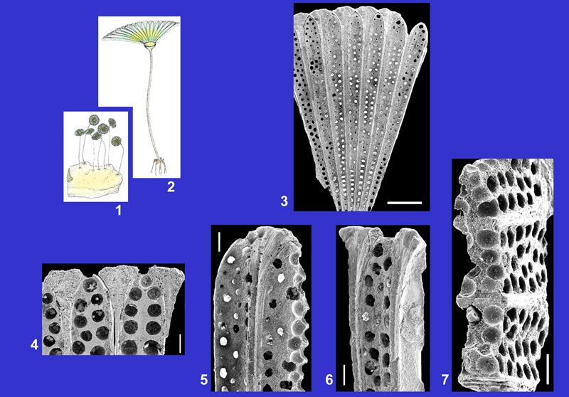
Dasycladales are unicellular green algae in existence since the Paleozoic
era. Dasycladales discovered in the Cenozoic strata of the French sedimentary basins
(Fig. 1 ![]() ) are noteworthy for the exceptional quality of their preservation. Although most fossil Dasycladales are known only in thin sections, the coatings of the Dasycladales in these basins, particularly of those in Lutetian beds, are easy to extract from sandy sediments and then are examined under the electron microscope. This method of investigation facilitates greatly the identification of the external and internal features of each species.
) are noteworthy for the exceptional quality of their preservation. Although most fossil Dasycladales are known only in thin sections, the coatings of the Dasycladales in these basins, particularly of those in Lutetian beds, are easy to extract from sandy sediments and then are examined under the electron microscope. This method of investigation facilitates greatly the identification of the external and internal features of each species.
Click on the image to enlarge it.
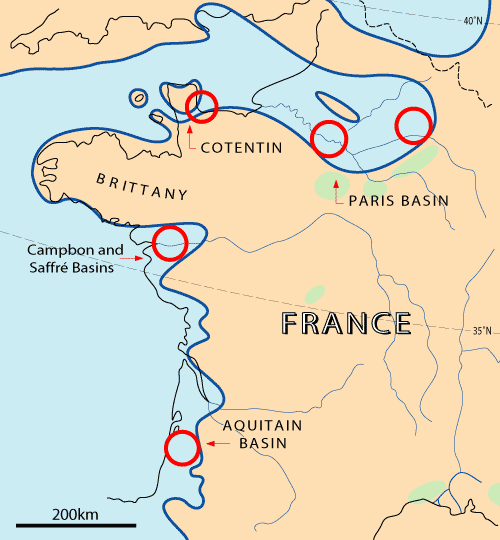
Figure 1: Paleogeographical map of France during Lutetian and main sites of discoveries (red circles).
Dasycladales flourished during the Lutetian because conditions
for their development were optimal during this epoch: hot climate and shallow marine water environments. The richest communities are composed of 13 to 18 species discovered in the Middle Lutetian sediments of the Paris Basin, in the Yvelines department (La Ferme de l'Orme, Grignon, Septeuil, Villiers-Saint-Frédéric) and in the Upper Lutetian sediments of Campbon and Saffré Basins, in the south of Brittany,
Loire-Atlantique department
(Fig. 1 ![]() ).
).
The preservation of the Dasycladales as fossils depends on the degree to which the original calcification covered living parts of the alga.
The extent of the calcification may vary greatly from one species to another of the same genus, sometimes even between individuals of a species. Of the Lutetian species illustrated in this photo-atlas, some have a calcification that envelops almost all the vegetative and reproductive organs (Neomeris larvarioides, Cymopolia armorica, Cy. elongata, Cy. turgescens, Zittelina rotunda), while in others calcification is restricted to a group of reproductive organs (cluster of gametophores in Carpenterella jonesi, whorl of tubular gametophores in Clypeina marginiporella, Cl. stelliformis) or to laterals (Belzungia terquemi, Thyrsoporella cancellata). In Neomeris, the calcification is particularly variable from one species to another: the two orders of laterals and the gametophores are strongly calcified in Neomeris larvarioides; the gametophores are completely calcified but the laterals only partially in N. filiformis, N. fragilis, N. pustulosa, N. radiata and N. reticulata; the laterals are uncalcified but the gametophores are strongly calcified in N. bipartita and N. limbata. To conclude, some species are known only as elongated isolated spicules (pluricystic gametophores of Acicularia acuminata, A. cornigera, A. pavantina), elongated associated spicules (pluricystic gametophores of Acicularia costulata, A. heberti, A. munieri) or sphaerical to ovoid isolated corpuscles (pluricystic gametophores in Frederica villiersi, gametophores without cysts in Dameryella tuberosa).

Click on the image to enlarge it.
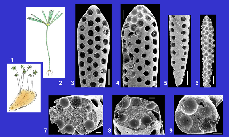
Figure 2: Main morphological features of the genera of which species are present in the Lutetian sediments of the French sedimentary basins.
Illustrated species:
- Acicularia acuminata , 1989
- Acicularia cornigera L. & J. , 1922
- Acicularia costulata , 1987
- Acicularia heberti L. & J. , 1922
- Acicularia munieri L. & J. , 1913
- Acicularia pavantina d', 1843
- Belzungia terquemi L. & J. , 1917
- Carpenterella jonesi L. & J. , 1922
- Clypeina marginiporella , 1845
- Clypeina stelliformis L. & J. , 1939
- Cymopolia armorica , 1987
- Cymopolia elongata (, 1825) , 1877
- Cymopolia turgescens , 1987
- Dameryella tuberosa & , 1998
- Frederica villiersi , 1966
- Neomeris bipartita , 1987
- Neomeris fercourtensis , 1980
- Neomeris filiformis (L. & J. , 1913) , 1927
- Neomeris fragilis (, 1825) L. & J. , 1913
- Neomeris larvarioides (L. & J. , 1913) , 1980
- Neomeris limbata (, 1822) , 1927
- Neomeris pustulosa L. & J. , 1917
- Neomeris radiata L. & J. , 1922
- Neomeris reticulata (, 1822) , 1927
- Thyrsoporella cancellata , 1872
- Zittelina dumasi (L. & J. , 1917) , 1927
- Zittelina elegans L. & J. , 1913
- Zittelina parisiensis (L. & J. , 1922) , 1927
- Zittelina rotunda (, 1980) , 1987
More informations and detailed descriptions concerning Lutetian Dasycladales of the French sedimentary basins are in the papers of L. & J. (1913, 1917, 1922) and (1978, 1980, 1987). Features of some Cenozoic species belonging to the genera Cymopolia, Neomeris and Acicularia are close to those of Recent species: the publications of (1968, 1969), (2006), and (1992) are useful for the interpretation of fossil specimens and for making comparative studies.
|
Acicularia acuminata , 1989 |
EOCENE | PRIABONIAN |
Click on the images below to enlarge the photomicrographs |
BARTONIAN | |
| LUTETIAN | ||
Description and more illustrations in: , 1987, p. 160, Pl. 10, fig. 14, Pl. 20, figs. 1-13; 1989, Pl. 1, figs. 7-11. |
YPRESIAN | |
| Plate 1 | ||
 |
||
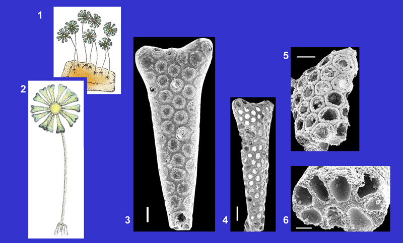
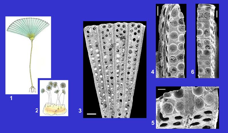
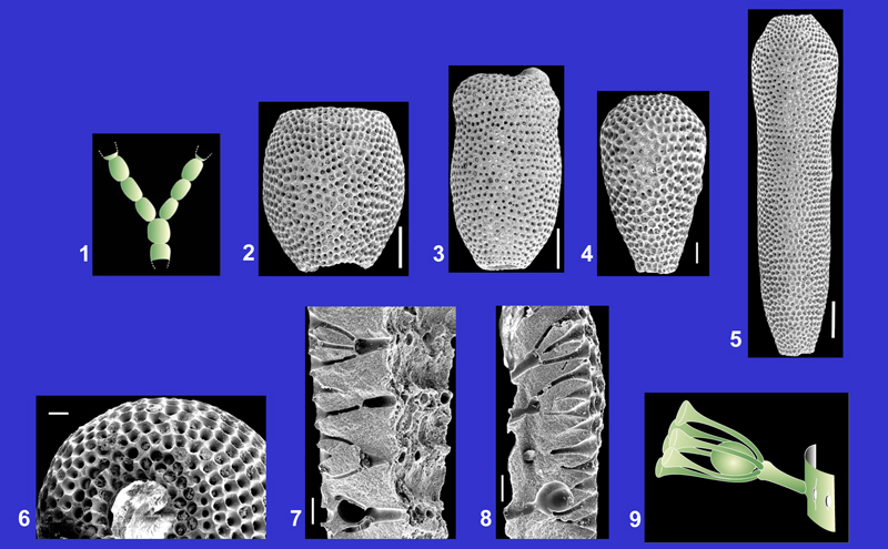
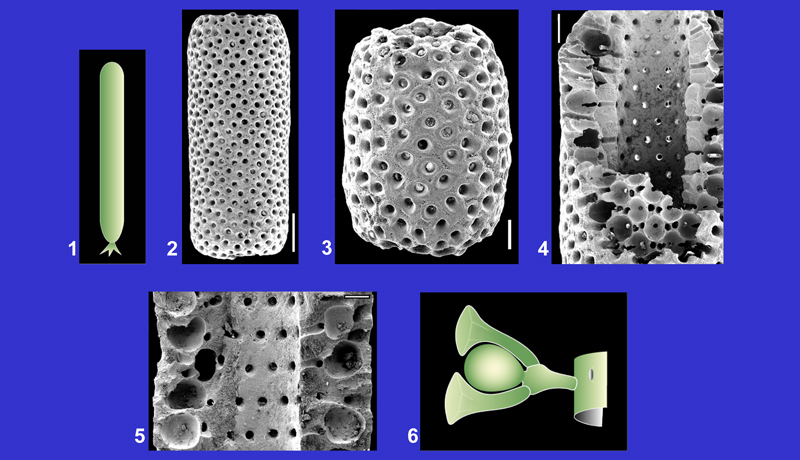
1. Hypothetical reconstruction of several living representatives. Drawing: Philippe .
2. A living representative, partially reconstructed. Drawing: Philippe .
3. External view of the outer part of a gametophore. Circular pores represent the location of the cysts. Scale: 100 μm.
4. External view of the outer part of a gametophore and section of the distal end showing cysts inside the wall. Scale: 50 μm.
5. Inner part of a gametophore. Scale: 100 μm.
6. Internal view of a gametophore with the location of cysts. Scale: 100 μm.
7. Transverse section of a gametophore showing the arrangement of the cysts at the periphery of the organ. Scale: 20 μm.
8. Idem.
9. Transverse section of the inner end of a gametophore. Scale: 20 μm.
Acicularia cornigera L. & J. , 1922 |
EOCENE | PRIABONIAN |
Click on the images below to enlarge the photomicrographs |
BARTONIAN | |
Upper Lutetian, Campbon (Loire-Atlantique), Brittany. |
LUTETIAN | |
Main references with description and illustrations: L. & J. , 1922, p. 24, Pl. 2, figs. 21-23; , 1987, p. 177, Pl. 15, figs. 1-12. |
YPRESIAN | |
| Plate 2 | ||
 |
||
1. Hypothetical reconstruction of several living representatives. Drawing: Philippe .
2. Reconstruction of a living representative. Drawing: Philippe .
3. External view of a gametophore. Middle Lutetian, Chaussy (Val-d'Oise), Paris Basin. Scale: 100 μm.
4. Idem. Middle Lutetian, Parnes-Montjavoult (Oise), Paris Basin. Scale: 200 μm.
5. Detail of the outer part of a weakly calcified gametophore. Middle Lutetian, Chaussy (Val-d'Oise), Paris Basin. Scale: 100 μm.
6. Transverse section of a gametophore with the location of cysts at the periphery of the organ. Middle Lutetian, Chaussy (Val-d'Oise), Paris Basin. Scale: 50 μm.
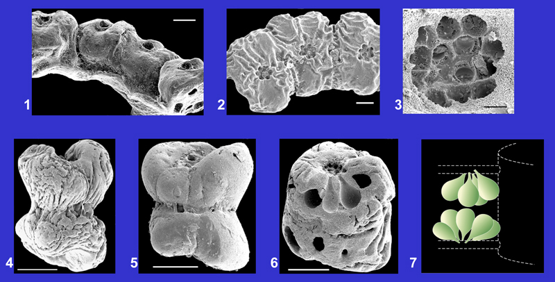
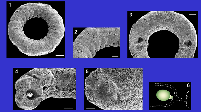
Acicularia costulata , 1987 |
EOCENE | PRIABONIAN |
Click on the images below to enlarge the photomicrographs |
BARTONIAN | |
Middle Lutetian, Thionville-sur-Opton (Eure), Paris Basin. |
LUTETIAN | |
Description and other illustrations: , 1987, p. 182, Pl. 16, figs. 1-10. |
YPRESIAN | |
| Plate 3 | ||
 |
||
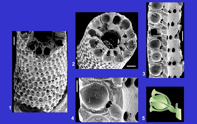
1. Hypothetical reconstruction of several living representatives. Drawing: Philippe .
2. A living representative, partially reconstructed. Drawing: Philippe .
3. Fragment of a reproductive disc composed of 5 gametophores. Scale: 200 μm.
4. Longitudinal section of a gametophore crossing the location of cysts. Scale: 100 μm.
5. Longitudinal section crossing the cysts (upper part of the photography) and typical aspect of the lateral wall of a gametophore (middle part of the photography). Scale: 100 μm.
6. Inner part of a reproductive disc and transverse sections of the proximal ends of several gametophores. Scale: 100 μm.
7. Transverse sections of 2 gametophores with the rounded cavities of cysts. Scale: 100 μm.
| EOCENE | PRIABONIAN | |
Click on the images below to enlarge the photomicrographs |
BARTONIAN | |
Middle Lutetian, Villiers-Saint-Frédéric (Yvelines), Paris Basin. |
LUTETIAN | |
Main references with description and illustrations: , 1987, p. 194, Pl. 9, fig. 16, Pl. 17, figs. 1-14. |
YPRESIAN | |
| Plate 4 | ||
 |
||
1. A living representative, partially reconstructed. Drawing: Philippe .
2. Hypothetical reconstruction of several living representatives. Drawing: Philippe
3. Fragment of a reproductive disc composed of 4 gametophores. Scale: 200 μm.
4. Longitudinal section of a gametophore crossing the cavities of cysts. Scale: 100 μm.
5-6. Transverse sections of gametophores crossing the rounded cavities of cysts. Scales: 50 μm (fig. 5) and 100 μm (fig. 6).
| EOCENE | PRIABONIAN | |
Click on the images below to enlarge the photomicrographs |
BARTONIAN | |
| LUTETIAN | ||
Main references with description and illustrations: L. & J. , 1913, p. 33, Pl. 3, figs. 15-19; 1922, p. 26, Pl. 2, fig. 34; , 1987, p. 217, Pl. 3, figs. 4-5, Pl. 5, fig. 3, Pl. 18, figs. 1-15. |
YPRESIAN | |
| Plate 5 | ||
 |
||
1. Hypothetical reconstruction of several living representatives. Drawing: Philippe .
2. A living representative, partially reconstructed. Drawing: Philippe .
3. Fragment of a reproductive disc composed of 7 gametophores. Middle Lutetian, Parnes (Oise), Paris Basin. Scale: 500 μm.
4. Detail of the outer parts of 2 gametophores separated by fan-shaped lamellae. Middle Lutetian, Ferme de l'Orme (Yvelines), Paris Basin. Scale: 100 μm.
5. External view of 2 gametophores and longitudinal section crossing the location of the cysts. Middle Lutetian, Damery (Marne), Paris Basin. Scale: 100 μm.
6. External view of a gametophore and lateral view of the lamellae separating the gametophores. Middle Lutetian, Damery (Marne), Paris Basin. Scale: 100 μm.
7. Transverse sections of 3 gametophores crossing the rounded cavities of cysts. Middle Lutetian, Villiers-Saint-Frédéric (Yvelines), Paris Basin. Scale: 100 μm.
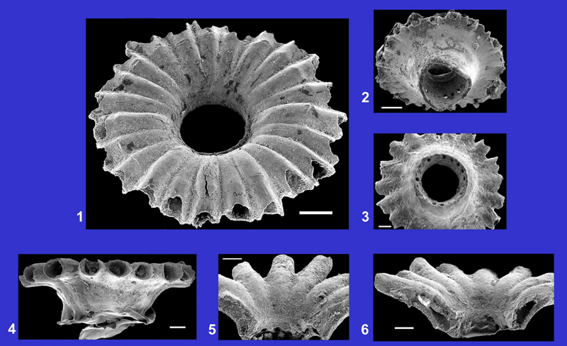
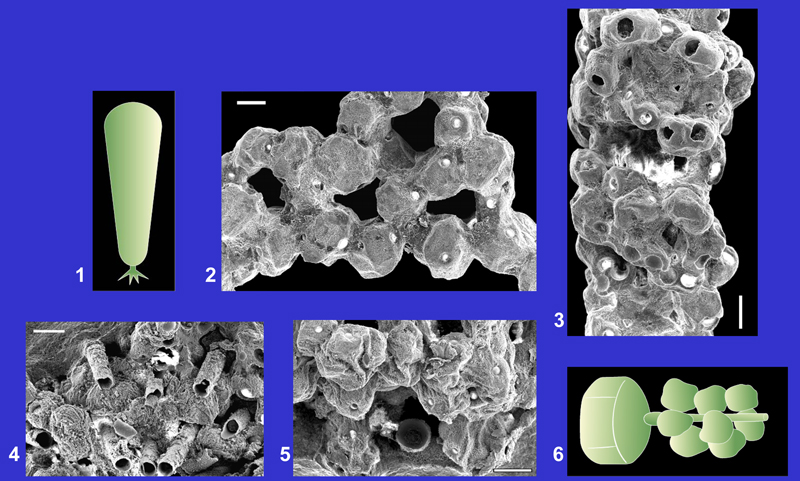
Acicularia pavantina d', 1843 |
EOCENE | PRIABONIAN |
Click on the images below to enlarge the photomicrographs |
BARTONIAN | |
|
LUTETIAN | |
Main references with description and illustrations: L. & J. , 1913, p. 32, Pl. 3, figs. 35-37; , 1987, p. 229, Pl. 3, fig. 6, Pl. 19, figs. 1-13. |
YPRESIAN | |
| Plate 6 | ||
 |
||
1. External view of a curved gametophore. Middle Lutetian, Villiers-Saint-Frédéric (Yvelines), Paris Basin. Scale: 500 μm.
2. External view of a straight gametophore. Middle Lutetian, La Ferme de l'Orme (Yvelines), Paris Basin. Scale: 200 μm.
3. Idem. Outer parts of the gametophores have 2 lobes more or less differenciated (compare figs. 1-3). Middle Lutetian, La Ferme de l'Orme (Yvelines), Paris Basin. Scale: 200 μm.
4. Longitudinal section of a hollow gametophore (lower part of the photo). Middle Lutetian, La Ferme de l'Orme (Yvelines), Paris Basin. Scale: 100 μm.
5. Transverse section of a gametophore. Cysts are located at the inner periphery of the gametophore. Middle Lutetian, La Ferme de l'Orme (Yvelines), Paris Basin. Scale: 100 μm.
6. Idem. This gametophore is almost entirely filled by the calcification. Middle Lutetian, La Ferme de l'Orme (Yvelines), Paris Basin. Scale: 100 μm.
7. Oblique section of a hollow gametophore. Middle Lutetian, La Ferme de l'Orme (Yvelines), Paris Basin. Scale: 100 μm.
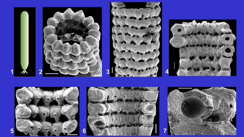
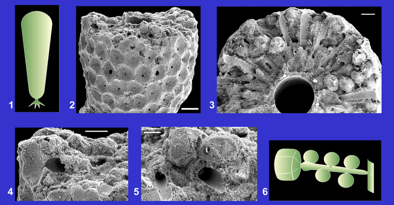
Belzungia terquemi L. & J. , 1917 |
EOCENE | PRIABONIAN |
Click on the images below to enlarge the photomicrographs |
BARTONIAN | |
|
LUTETIAN | |
Main references with description and illustrations: L. & J. , 1917, p. 370, Pl. 14, figs. 13-17; , 1966, p. 137, Pl. 1, figs. 7-16; , 1987, p. 263, Pl. 10, figs. 16-17, Pl. 42, figs. 1-11. |
YPRESIAN | |
| PALEOCENE | ||
| Plate 7 | ||
 |
||
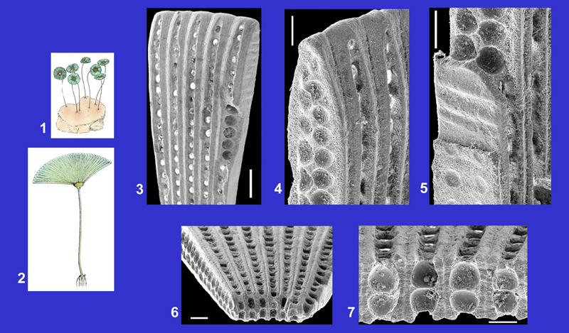
1. Hypothetical general shape of the thallus.
2. External general view of the sheath. Upper Lutetian, Campbon (Loire-Atlantique), Brittany. Scale: 500 μm.
3. Detail of the external surface. The irregularly arranged pores represent the location of the last order lateral segments. Upper Lutetian, Le Bois-Gouët (Loire-Atlantique), Brittany. Scale: 100 μm.
4. External general view of a slightly worn specimen. Upper Lutetian, Le Bois-Gouët (Loire-Atlantique), Brittany. Scale: 500 μm.
5. Detail of a worn surface. The pores correspond to the location of the three orders of lateral segments. Upper Lutetian, Le Bois-Gouët (Loire-Atlantique), Brittany. Scale: 100 μm.
6. Transverse section of the calcareous sheath. Each primary lateral segment branches several times inside the wall. Upper Lutetian, Campbon (Loire-Atlantique), Brittany. Scale: 200 μm.
7. Internal surface of the sheath (left side) with the location of the primary lateral segments (large pores) and longitudinal section (right side) with divisions of the lateral segments (canals). Upper Lutetian, Campbon (Loire-Atlantique), Brittany. Scale: 100 μm.
| EOCENE | PRIABONIAN | |
Click on the images below to enlarge the photomicrographs |
BARTONIAN | |
|
LUTETIAN | |
Main references with description and illustrations: L. & J. , 1922, p. 20, Pl. 1, figs. 77-80; , 1980, Pl. 22, figs. 12-15; 1987, p. 270, Pl. 30, figs. 1-13. |
YPRESIAN | |
| PALEOCENE | ||
| Plate 8 | ||
 |
||
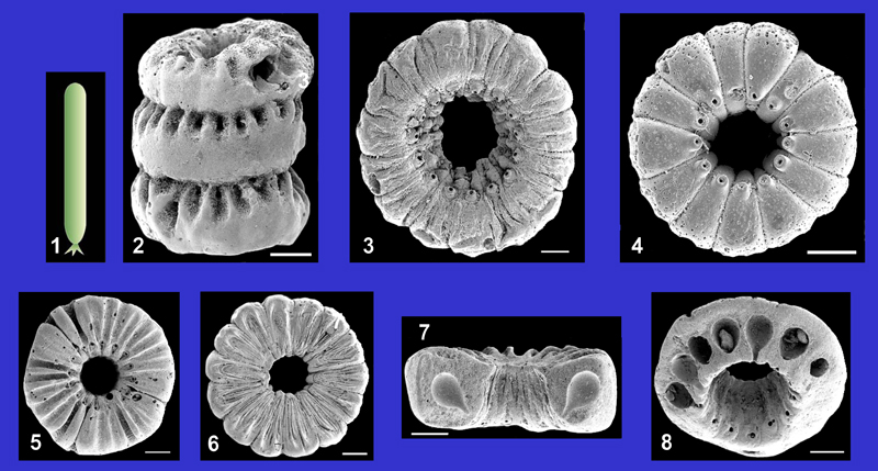
1. Fragment of a ring composed of numerous associated corpuscles, each containing a cluster of gametophores. Middle Lutetian, Grignon (Yvelines), Paris Basin. Scale: 100 μm.
2. Fragment of a ring composed of 3 corpuscles. Upper Lutetian, Campbon (Loire-Atlantique), Brittany. Scale: 50 μm.
3. Group of pores representing the location of the peduncles of the gametophores at the surface of a corpuscle. Middle Lutetian, Septeuil (Yvelines), Paris Basin. Scale: 10 μm.
4-5. Associations of 2 corpuscles with different types of external surfaces. Middle Lutetian, Septeuil (Yvelines), Paris Basin. Scale: 100 μm.
6. Pedunculate gametophores inside a corpuscle. Middle Lutetian, Septeuil (Yvelines), Paris Basin. Scale: 100 μm.
7. Reconstruction of 2 clusters of gametophores located along the laterals. Drawing: Robert .
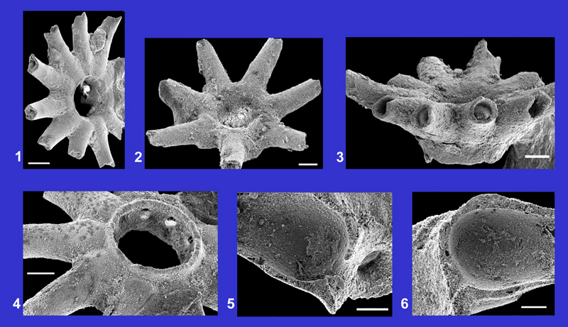
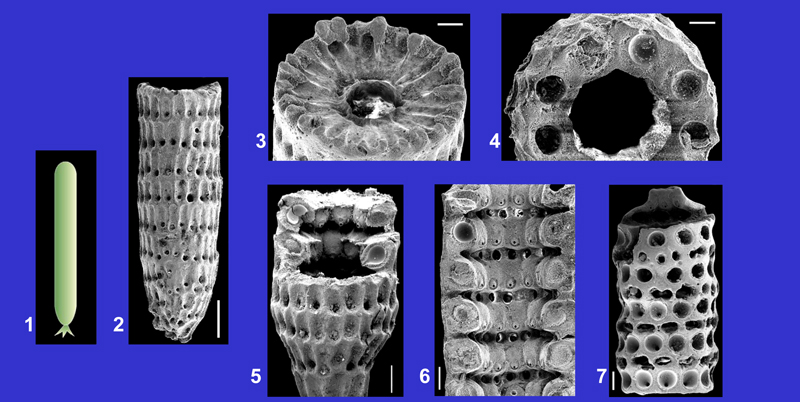
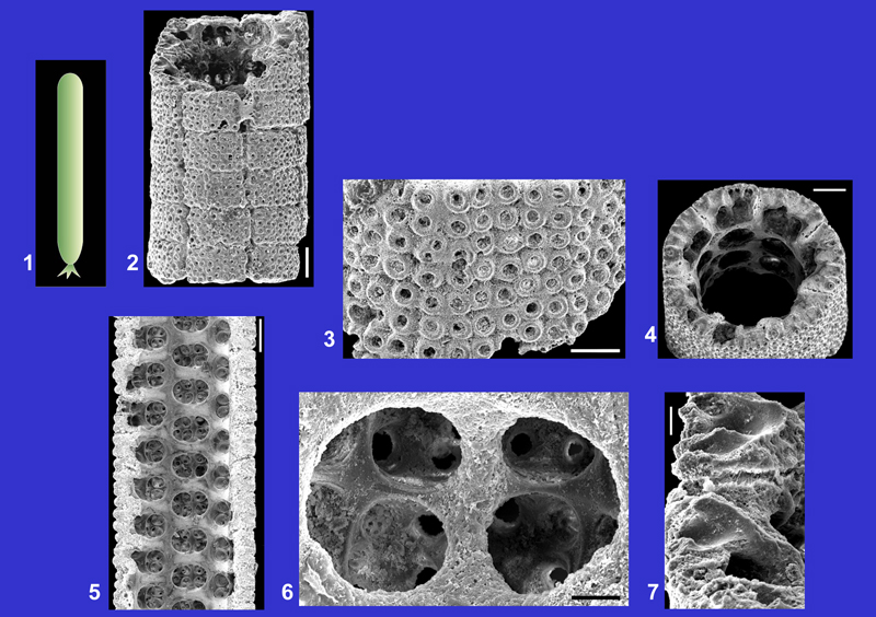
Clypeina marginiporella , 1845 |
OLIGOCENE | STAMPIAN |
Click on the images below to enlarge the photomicrographs |
EOCENE | PRIABONIAN |
|
|
BARTONIAN | |
Middle Lutetian sediments, Chambors (Oise), Paris Basin. |
LUTETIAN | |
Main references with description and illustrations: L. & J. , 1913, p. 34, Pl. 3, figs. 20-25; 1922, p. 27, Pl. 2, figs. 45-46; , 1987, p. 244, Pl. 26, figs. 1-10. |
YPRESIAN | |
| Plate 9 | ||
 |
||
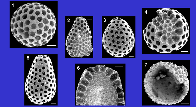
1. Upper side of a whorl of gametophores. Scale: 200 μm.
2-3. Lower side of whorls of gametophores. Scales: 200 μm (fig. 2) and 100 μm (fig. 3).
4. Lateral view of the gametophores, partially preserved. Scale: 100 μm.
5-6. Longitudinal sections of gametophores and habit of the axial cavity. Scales: 100 μm.
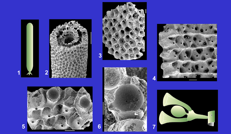
Clypeina stelliformis L. & J. , 1939 |
EOCENE | PRIABONIAN |
Click on the images below to enlarge the photomicrographs |
BARTONIAN | |
| LUTETIAN | ||
Main references with description and illustrations: L. & J. , 1913 (Cl. digitata), p. 35, Pl. 3, figs. 26-31; , 1987, p. 249, Pl. 27, figs. 1-11. |
YPRESIAN | |
| Plate 10 | ||
 |
||
1. Upper side of a whorl of tubular gametophores. Middle Lutetian, Chambors (Oise), Paris Basin. Scale: 200 μm.
2. Upper side of a whorl of tubular gametophores. Middle Lutetian, Chaumont-en-Vexin (Oise), Paris Basin. Scale: 100 μm.
3. Lateral view of the gametophores, partially preserved. Middle Lutetian, Chambors (Oise), Paris Basin. Scale: 100 μm.
4. Lower side of a whorl of gametophores. A row of pores is located on the circular wall of the axial cavity, representing the proximal ends of the gametophores. Middle Lutetian, Chaumont-en-Vexin (Oise), Paris Basin. Scale: 100 μm.
5-6. Longitudinal sections of the lower parts of gametophores. Middle Lutetian, Chambors (Oise), Paris Basin. Scales: 50 μm.
Cymopolia armorica , 1987 |
EOCENE | PRIABONIAN |
Click on the images below to enlarge the photomicrographs |
BARTONIAN | |
Upper Lutetian, Le Bois-Gouët (Loire-Atlantique), Brittany. |
LUTETIAN | |
Description and other illustrations: , 1987, p. 276, Pl. 37, figs. 7-12. |
YPRESIAN | |
| Plate 11 | ||
 |
||
1. Lower part of the picture: external surface of the sheath. The large pores represent the location of the upper ends of the secondary lateral segments. Upper part of the picture: internal structure of the wall. Large circular cavities represent the transverse sections of the gametophores, surrounded by numerous small pores (transverse sections of the secondary segments). Paratype (cf. , 1987, Pl. 37, fig. 12). Scale: 100 μm.
2. Left side: longitudinal section of the wall. Each primary lateral segment branches into several secondary segments surrounding an ovoid gametophore. Right side: internal surface of the sheath. Small circular pores are the proximal ends of primary lateral segments, arranged in whorls. Paratype (cf. , 1987, fig. 8). Scale: 200 μm.
3. Reconstruction of 2 orders of laterals and of a gametophore. Drawing: Philippe .
Cymopolia elongata (, 1825) , 1877 |
MIOCENE |
Click on the images below to enlarge the photomicrographs |
OLIGOCENE |
Middle Lutetian, Grignon (Yvelines), Paris Basin. |
EOCENE |
Main references with description and illustrations: L. & J. , 1913, p. 10, Pl. 1, figs. 1-12; 1922, p. 8, Pl. 1, figs. 1-7, 9-11; , 1980, Pl. 1, figs. 1-12, Pl. 3, figs. 6-9. |
PALEOCENE |
| Plate 12 | |
 |
|
1. Schematic reconstruction of the thallus, composed of superposed segments.
2-5. External views of 4 specimens, displaying the great morphological variability of the articles. Scales: 500 μm (figs. 2-3, 5) and 200 μm (fig. 4).
6. Upper end of a segment and detail of the external surface. The alveols represent the enlarged distal ends of the secondary lateral segments. Scale: 100 μm.
7-8. Longitudinal sections of the wall with the 2 orders of segments (canals) and the gametophores (rounded cavities). Scales: 100 μm.
9. Reconstruction of 2 orders of segments and of a gametophore. Drawing: Robert .
Cymopolia turgescens , 1987 |
EOCENE | PRIABONIAN |
Click on the images below to enlarge the photomicrographs |
BARTONIAN | |
| LUTETIAN | ||
Description and other illustrations: , 1987, p. 285, Pl. 10, figs. 9-11, Pl. 37, figs. 1-6. |
YPRESIAN | |
| Plate 13 | ||
 |
||
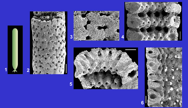
1. External view. The surface is composed of alveols representing the enlarged distal ends of the secondary lateral segments. Upper Lutetian, Le Bois-Gouët (Loire-Atlantique), Brittany. Scale: 100 μm.
2. Transverse section with the 2 orders of lateral segments (canals) and the gametophores (large rounded cavities). The central cavity is narrow. Upper Lutetian, Pierre-Aiguë (Loire-Atlantique), Brittany. Scale: 100 μm.
3. Longitudinal section of the wall crossing the 2 orders of lateral segments and the gametophores. Upper Lutetian, Le Bois-Gouët (Loire-Atlantique), Brittany. Scale: 100 μm.
4. Detail of the wall with 2 orders of lateral segments and a gametophore. Upper Lutetian, Le Bois-Gouët (Loire-Atlantique), Brittany. Scale: 50 μm.
5. Reconstruction of a primary lateral segment bearing several secondary segments and a gametophore. Drawing: Philippe .
Dameryella tuberosa & , 1998 |
EOCENE | PRIABONIAN |
Click on the images below to enlarge the photomicrographs |
BARTONIAN | |
| LUTETIAN | ||
Description and other illustrations: & , 1998, p. 20, Pl. 1, figs. 1-12. |
YPRESIAN | |
| Plate 14 | ||
 |
||
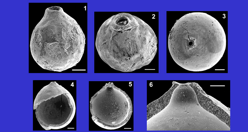
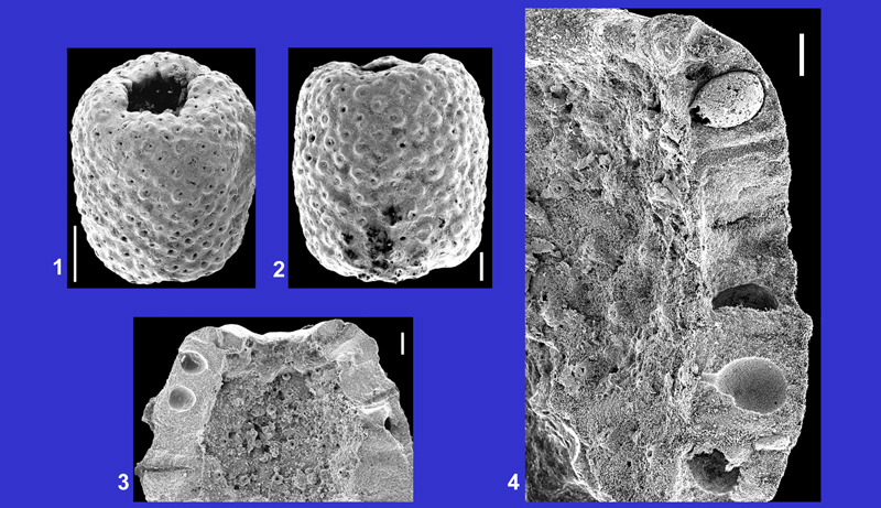
1. External view. Middle Lutetian, Grignon (Yvelines), Paris Basin. Scale: 100 μm.
2. External view. Middle Lutetian, Damery (Marne), Paris Basin. Scale: 50 μm.
3. External view. Middle Lutetian, Grignon (Yvelines), Paris Basin. Scale: 100 μm.
4. External and internal views. Middle Lutetian, Grignon (Yvelines), Paris Basin. Scale: 100 μm.
5. Internal view. Middle Lutetian, Grignon (Yvelines), Paris Basin. Scale: 100 μm.
6. Detail of the opening. Middle Lutetian, Grignon (Yvelines), Paris basin. Scale: 50 μm.
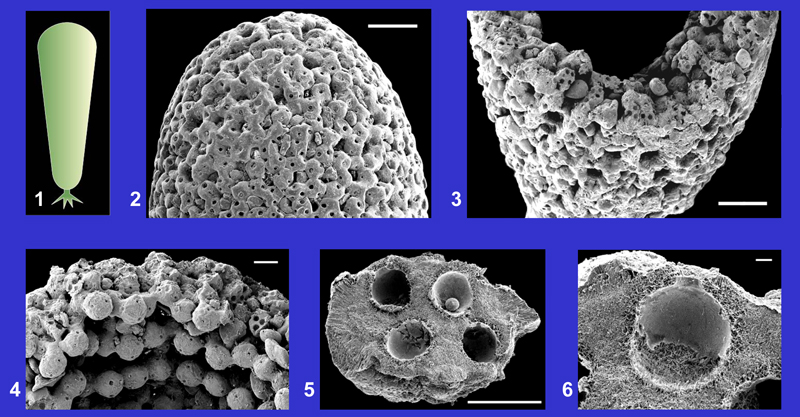
Frederica villiersi , 1966 |
EOCENE | PRIABONIAN |
Click on the images below to enlarge the photomicrographs |
BARTONIAN | |
| LUTETIAN | ||
Main references with description and illustrations: , 1966, p. 907, Pl. 39, figs. 1-8, 11-13 ; , 1987, Pl. 41, figs. 1-12. |
YPRESIAN | |
| Plate 15 | ||
 |
||
1. External view of a sphaerical corpuscle. Middle Lutetian, Villiers-Saint-Frédéric (Yvelines), Paris Basin. Scale: 100 μm.
3 & 5. External views of two ovoid specimens. Upper Lutetian, Montchauvet (Yvelines), Paris Basin. Scales: 100 μm.
2, 4 & 6. Internal views displaying the location of the cysts at the periphery of the corpuscles. Upper Lutetian, Montchauvet (Yvelines), Paris Basin. Scales: 100 μm.
7. Internal view of a hollow specimen. Middle Lutetian, Villiers-Saint-Frédéric (Yvelines), Paris Basin. Scale: 100 μm.
Neomeris bipartita , 1987 |
EOCENE | PRIABONIAN |
Click on the images below to enlarge the photomicrographs |
BARTONIAN | |
Lower Lutetian, Boisgeloup (Mont-de-Magny, Eure), Paris Basin. |
LUTETIAN | |
Description and other illustrations: , 1987, p. 319, Pl. 32, figs. 6-13. |
YPRESIAN | |
| Plate 16 | ||
 |
||
1. Isolated ring representing the calcification of a circular row of gametophores. Lateral segments are not calcified. Scale: 200 μm.
2. Detail of the internal part of a ring. Pores correspond to the location of the basal ends of gametophores. Scale: 100 μm.
3. Transverse section of a ring crossing several gametophores. Scale: 100 μm.
4-5. Longitudinal sections crossing 2 gametophores. Scales: 100 μm (fig. 4) and 50 μm (fig. 5).
6. Reconstruction of the gametophore and hypothetical reconstruction of the orders of lateral segments. Drawing: Alain .
Neomeris fercourtensis , 1980 |
EOCENE | PRIABONIAN |
Click on the images below to enlarge the photomicrographs |
BARTONIAN | |
Middle Lutetian, Fercourt (Oise), Paris Basin. |
LUTETIAN | |
Description and other illustrations: , 1980, Pl. 7, figs. 7-12, Pl. 10, figs. 9-10, Pl. 11, figs. 7-8, Pl. 12, fig. 6. |
YPRESIAN | |
| Plate 17 | ||
 |
||
1. Schematic reconstruction of the general shape of the thallus.
2. External view with the typical alveolated aspect of the surface. Scale: 100 μm.
3-4. Internal surfaces and longitudinal sections of the wall across some gametophores and secondary lateral segments. Primary lateral segments are not calcified. Scales: 100 μm.
Neomeris filiformis (L. & J. , 1913) , 1927 |
EOCENE | PRIABONIAN |
Click on the images below to enlarge the photomicrographs |
BARTONIAN | |
| LUTETIAN | ||
Main references with description and illustrations: L. & J. , 1913, p. 17, Pl. 1, figs. 52-53; , 1980, Pl. 16, figs. 1-9, Pl. 18, figs. 5-6, Pl. 19, figs. 8-9, Pl. 20, fig. 5. |
YPRESIAN | |
| Plate 18 | ||
 |
||
1. Schematic reconstruction of the general shape of the thallus.
2. External view with the typical alveolated aspect of the surface. Middle Lutetian, Chaussy (Oise), Paris Basin. Scale: 200 μm.
3. Transverse section with the location of the secondary lateral segments (primary segments are not calcified). Middle Lutetian, Damery (Marne), Paris Basin. Scale: 100 μm.
4. Transverse section with the location of the gametophores. The central cavity is narrow. Middle Lutetian, Chaussy (Oise), Paris Basin. Scale: 50 μm.
5. Longitudinal section with sections of the gametophores and external view of the basal end of the sheath. Middle Lutetian, Chaussy (Oise), Paris Basin. Scale: 100 μm.
6. Longitudinal section and internal surface presenting alternate swellings (envelopes of gametophores) and circular furrows (location of secondary lateral segments: rounded pores). Middle Lutetian, Chaussy (Oise), Paris Basin. Scale: 100 μm.
7. Tangential section with alternation of rows of large rounded cavities (gametophores) and rows of small pores (secondary segments partially calcified). Middle Lutetian, Damery (Marne), Paris Basin. Scale: 100 μm.
Neomeris fragilis (, 1825) L. & J. , 1913 |
EOCENE | PRIABONIAN |
Click on the images below to enlarge the photomicrographs |
BARTONIAN | |
Middle Lutetian, Grignon (Yvelines), Paris Basin. |
LUTETIAN | |
Main references with description and illustrations: L. & J. , 1913, p. 20, Pl. 2, figs. 15-19; , 1980, Pl. 6, figs. 1-9, Pl. 10, figs. 7-8, Pl. 11, fig. 9, Pl. 12, figs. 4-5; 1987, Pl. 1, figs. 1-2. |
YPRESIAN | |
| Plate 19 | ||
 |
||
1. Schematic reconstruction of the general shape of the thallus.
2. External view with the alveolated surface (calcification of distal ends of the secondary lateral segments) and transverse section showing the calcification of the stem. Scale: 500 μm.
3. Detail of the external surface. Scale: 100 μm.
4. Internal surface with the calcification of the gametophores (small circular pores represent the location of the peduncles). Very irregular pores correspond to the location of the secondary lateral segments, very partially and irregularly calcified. Scale: 100 μm.
5. Detail of a transverse section with 2 gametophores. Scale: 100 μm.
6. A gametophore with its short peduncle. Scale: 50 μm.
7. Reconstruction of a gametophore and hypothetical reconstruction of the lateral segments (lateral segments are almost not calcified in this species). Drawing: Robert .
| EOCENE | PRIABONIAN | |
Click on the images below to enlarge the photomicrographs |
BARTONIAN | |
Upper Lutetian, Le Bois-Gouët (Loire-Atlantique), Brittany. |
LUTETIAN | |
Main references with description and illustrations: L. & J. , 1913, p. 13, Pl. 1, figs. 41-42; , 1980, Pl. 8, figs. 1-10; 1987, Pl. 10, figs. 1-3, Pl. 33, figs. 1-13. |
YPRESIAN | |
| Plate 20 | ||
 |
||
1. Schematic reconstruction of the general shape of the thallus.
2-3. Different aspects of the sheath. Pores are the location of the secondary lateral segments. Scales: 500 μm (fig. 2) and 200 μm (fig. 3).
4. Internal aspect and longitudinal section of the wall with the 2 orders of laterals and the gametophores. Scale: 200 μm.
5. Longitudinal section. Calcification surrounds the laterals and the gametophores. Scale: 100 μm.
6. Reconstruction of a gametophore surrounded by the 2 orders of laterals. Drawing: Robert .
Neomeris limbata (, 1822) , 1927 |
EOCENE | PRIABONIAN |
Click on the images below to enlarge the photomicrographs |
BARTONIAN | |
| LUTETIAN | ||
Main references with description and illustrations: L. & J. , 1913, p. 14, Pl. 1, figs. 25-35; , 1980, Pl. 14, figs. 1-12, Pl. 18, figs. 9-11, Pl. 19, figs. 4-5, 10. |
YPRESIAN | |
| PALEOCENE | THANETIAN | |
| Plate 21 | ||
 |
||
1. Schematic reconstruction of the general shape of the thallus.
2. Superposed rings corresponding to the calcification of 3 circular rows of gametophores. Pores between rings are the location of the secondary lateral segments, almost not preserved. Middle Lutetian, Saint-Lubin-de-la-Haye (Eure), Paris Basin. Scale: 100 μm.
3-5. Different aspects of the ornamentation on the lower side of rings. Each unit corresponds to the calcification of a gametophore. Pores of the inner periphery are the location of peduncles. 3: Middle Lutetian, La Ferme de l'Orme (Yvelines), Paris Basin. 4: Upper Lutetian, Le Bois-Gouët (Loire-Atlantique), Brittany. 5: Middle Lutetian, Saint-Lubin-de-la-Haye (Eure), Paris Basin. Scales: 100 μm.
6. Upper side of a ring. Middle Lutetian, Saint-Lubin-de-la-Haye (Eure), Paris Basin. Scale: 100 μm.
7. Longitudinal section of a ring crossing 2 gametophores. Middle Lutetian, Saint-Lubin-de-la-Haye (Eure), Paris Basin. Scale: 100 μm.
8. Circular row of gametophores inside a ring. Middle Lutetian, La Ferme de l'Orme (Yvelines), Paris Basin. Scale: 100 μm.
Neomeris pustulosa L. & J. , 1917 |
EOCENE | PRIABONIAN |
Click on the images below to enlarge the photomicrographs |
BARTONIAN | |
Upper Lutetian, Le Bois-Gouët (Loire-Atlantique), Brittany. |
LUTETIAN | |
Description and other illustrations: L. & J. , 1917, p. 366, Pl. 14, figs. 4-6; , 1987, Pl. 10, figs. 4-5; Pl. 34, figs. 1-12. |
YPRESIAN | |
| Plate 22 | ||
 |
||
1-2. External aspects of articles. Scales: 500 μm (fig. 1) and 200 μm (fig. 2).
3-4. Longitudinal sections of articles crossing gametophores and rare secondary lateral segments. Scales: 100 μm.
Neomeris radiata L. & J. , 1922 |
EOCENE | PRIABONIAN |
Click on the images below to enlarge the photomicrographs |
BARTONIAN | |
Middle Lutetian, Liancourt-Saint-Pierre (Oise), Paris Basin (except fig. 2: Fercourt (Oise)). |
LUTETIAN | |
Main references with description and illustrations: L. & J. , 1922, p. 14, Pl. 1, figs. 33-37; , 1980, Pl. 7, figs. 1-6, Pl. 10, figs. 5-6, Pl. 11, figs. 5-6, Pl. 12, fig. 3. |
YPRESIAN | |
| Plate 23 | ||
 |
||
1. Schematic reconstruction of the general shape of the thallus.
2. External aspect of the sheath. Pores are the location of the secondary lateral segments. Scale: 200 μm.
3. Detail of the external surface. Each unit is the calcification of a gametophore. The wall surface is perforated by numerous small pores of irregular shape. Large pores correspond to the location of secondary lateral segments, partially calcified. Scale: 100 μm.
4. Internal surface with superposed rings, each corresponding to the calcification of a circular row of gametophores. Secondary laterals are located between these rings. Scale: 100 μm.
5. Two superposed rings. Rows of small rounded pores, located at the inner periphery of the rings, are the proximal ends of the peduncles of gametophores. Scale: 100 μm.
6. Longitudinal section crossing gametophores and some secondary lateral segments. Scale: 100 μm.
Neomeris reticulata (, 1822) , 1927 |
EOCENE | PRIABONIAN |
Click on the images below to enlarge the photomicrographs |
BARTONIAN | |
Middle Lutetian, Grignon (figs. 3-4, 6) and La Ferme de l'Orme (figs. 2, 5, 7), Yvelines, Paris Basin. |
LUTETIAN | |
Main references with description and illustrations: L. & J. , 1913, p. 14, Pl. 1, figs. 36-40; , 1980, Pl. 15, figs. 1-9, Pl. 18, figs. 3-4, Pl. 19, figs. 6-7, Pl. 20, figs. 3-4. |
YPRESIAN | |
| Plate 24 | ||
 |
||
1. Schematic reconstruction of the general shape of the thallus.
2. External aspect of the sheath. The calcified envelopes of the gametophores look like associated rounded corpuscles. Scale: 200 μm.
3. External aspect of a strongly calcified specimen. Pores are the location of the secondary lateral segments, partially calcified. Scale: 100 μm.
4. Internal surface of the sheath. Each ring corresponds to the calcification of a circular row of gametophores. Rows of gametophores are irregularly calcified. Secondary laterals are located between the rings. Scale: 100 μm.
5. Longitudinal section and internal surface of a weakly calcified specimen. Scale: 100 μm.
6. Longitudinal section, crossing gametophores and secondary laterals, and internal surface of a strongly calcified specimen. Scale: 100 μm.
7. Detail of the longitudinal section crossing a gametophore. Scale: 50 μm.
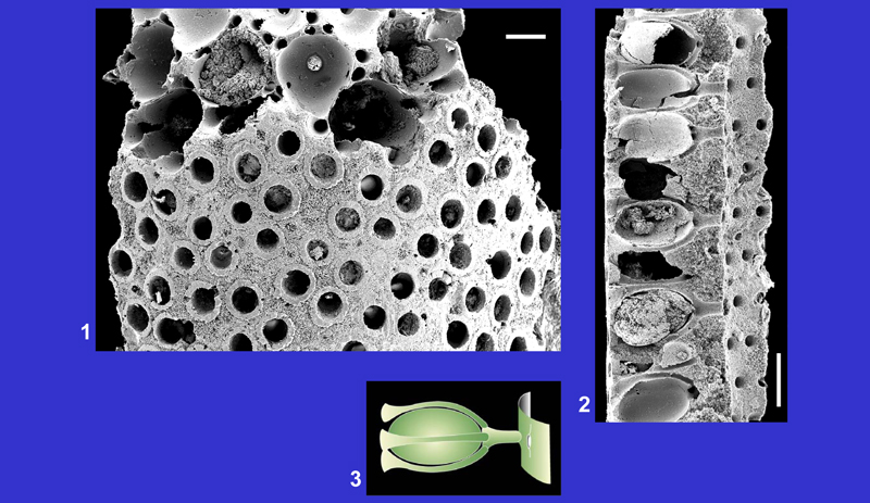
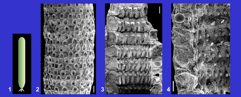
Thyrsoporella cancellata , 1872 |
EOCENE | PRIABONIAN |
Click on the images below to enlarge the photomicrographs |
BARTONIAN | |
| LUTETIAN | ||
Main references with description and illustrations: L. & J. , 1913, p. 37, Pl. 3, figs. 32-34; , 1966, p. 136, Pl. 1, figs. 1-6; , 1980, Pl. 25, figs. 4-7. |
YPRESIAN | |
| Plate 25 | ||
 |
||
1. Schematic reconstruction of the general shape of the thallus.
2. External aspect of the sheath. Each plate has 32 pores which correspond to the last order laterals, with 4 horizontal and 8 vertical rows of pores. Middle Lutetian, Châteaurouge (Oise), Paris Basin. Scale: 100 μm.
3. Detail of the external surface with the rows of the last order laterals. Middle Lutetian, Fercourt (Oise), Paris Basin. Scale: 50 μm.
4. Transverse section showing the large main axis and the division of the laterals inside the wall. Middle Lutetian, Fercourt (Oise), Paris Basin. Scale: 100 μm.
5. Longitudinal section. Middle Lutetian, Fercourt (Oise), Paris Basin. Scale: 100 μm.
6. Detail of the internal surface. One primary lateral segment (large oval pore) branches into 2 secondary laterals separated by a vertical septum. Middle Lutetian, Châteaurouge (Oise), Paris Basin. Scale: 20 μm.
7. Oblique section crossing several orders of laterals inside the wall. Middle Lutetian, Châteaurouge (Oise), Paris Basin. Scale: 20 μm.
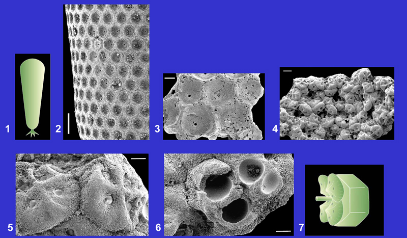
Zittelina dumasi (L. & J. , 1917) , 1927 |
EOCENE | PRIABONIAN |
Click on the images below to enlarge the photomicrographs |
BARTONIAN | |
Upper Lutetian, Le Bois-Gouët (Loire-Atlantique), Brittany. |
LUTETIAN | |
Main references with description and illustrations: L. & J. , 1917, p. 369, Pl. 14, figs. 11-12; , 1987, Pl. 10, figs. 18-19, Pl. 39, figs. 9-14; 1989, Pl. 1, figs. 14-15. |
YPRESIAN | |
| Plate 26 | ||
 |
||
1. Schematic reconstruction of the general shape of the thallus.
2. Top of the sheath. Scale: 500 μm.
3. Section of the wall composed of numerous gametophores closely attached by their envelopes. Small rounded perforations of the envelopes correspond to the location of cysts. Scale: 500 μm.
4. Detail of the wall displaying the irregular arrangement of the gametophores. Scale: 200 μm.
5. Section of a gametophore crossing the rounded cavities of the cysts. Scale: 100 μm.
6. Detail of the calcification surrounding a cyst. Scale: 10 μm.
Zittelina elegans L. & J. , 1913 |
EOCENE | PRIABONIAN |
Click on the images below to enlarge the photomicrographs |
BARTONIAN | |
| LUTETIAN | ||
Main references with description and illustrations: L. & J. , 1913, p. 27, Pl. 3, figs. 5-6; , 1980, Pl. 24, figs. 7-8, 10-12; 1987, Pl. 9, fig. 8, Pl. 39, figs. 1-3. |
YPRESIAN | |
| Plate 27 | ||
 |
||
1. Schematic reconstruction of the general shape of the thallus.
2. Alveolate external surface of the sheath. Middle Lutetian, Grignon (Yvelines). Scale: 500 μm.
3. Detail of the external surface. Each alveole is the calcification of the enlarged distal end of a lateral segment. Middle Lutetian, Villiers-Saint-Frédéric (Yvelines). Scale: 100 μm.
4. Internal surface of the sheath composed of several groups of gametophores surrounding the upper part of the laterals. Middle Lutetian, La Ferme de l'Orme (Yvelines). Scale: 100 μm.
5. Detail of the internal surface with 2 gametophores. Middle Lutetian, La Ferme de l'Orme (Yvelines). Scale: 30 μm.
6. Internal aspect of a gametophore. Rounded cavities correspond to the location of cysts. Middle Lutetian, La Ferme de l'Orme (Yvelines). Scale: 30 μm.
7. Hypothetical reconstruction of the upper end of a lateral bearing 4 gametophores. Drawing: Robert .
Zittelina parisiensis (L. & J. , 1922) , 1927 |
EOCENE | PRIABONIAN |
Click on the images below to enlarge the photomicrographs |
BARTONIAN | |
Middle Lutetian , La Ferme de l'Orme (Yvelines) (figs. 2-3) and Gentilly (Val-de-Marne) (figs. 4-5), Paris Basin. |
LUTETIAN | |
Main references with description and illustrations: L. & J. , 1922, p. 18, Pl. 1, figs. 84-87; , 1980, Pl. 23, figs. 9-13; 1987, Pl. 39, figs. 4-8. |
YPRESIAN | |
| Plate 28 | ||
 |
||
1. Schematic reconstruction of the general shape of the thallus.
2. The wall is composed of numerous gametophores with calcareous envelopes tied together. Laterals are not preserved. Scale: 100 μm.
3. Internal aspect of the wall with section of some gametophores displaying the location of cysts (small rounded cavities). Laterals are not preserved. Scale: 100 μm.
4. Exceptionally preserved laterals (tubes) surrounded by the gametophores. Scale: 100 μm.
5. Group of gametophores around the upper end of a lateral (transverse section of the lateral at the lower part of the photography). Scale: 100 μm.
6. Reconstruction of the upper end of a lateral surrounded by the gametophores. Drawing: Robert .
| EOCENE | PRIABONIAN | |
Click on the images below to enlarge the photomicrographs |
BARTONIAN | |
Middle Lutetian, La Ferme de l'Orme (Yvelines), Paris Basin. |
LUTETIAN | |
Description and other illustrations: , 1980, Pl. 24, figs. 1-6, 9; 1987, Pl. 9, fig. 9. |
YPRESIAN | |
| Plate 29 | ||
 |
||
1. Schematic reconstruction of the general shape of the thallus.
2. External aspect of the lower part of the sheath. The cortical layer is composed of alveoles (enlarged distal ends of the laterals). Scale: 200 μm.
3. Transverse section. Only one order of laterals bearing gametophores with cysts inside. Scale: 100 μm.
4. Detail of the wall showing the longitudinal section of the upper part of a lateral with its enlarged distal end (left side of the photography). Scale: 100 μm.
5. Detail of the wall showing 2 laterals surrounded by gametophores. Scale: 50 μm.
6. Reconstruction of one lateral bearing several gametophores. Drawing: Robert .
The microphotographs were taken at the University of Nantes, with the practical help of Alain . I am indebted to Alain for the realization of the slides and to Robert and Philippe for the drawings. All my particular thanks to Bruno who made this publication possible and to Ioan I. and Filippo for their help in improving the manuscript.
Selected publications containing descriptions and illustrations of Lutetian species from French Cenozoic basins:
S. (1966).- Algues Dasycladacées du Lutétien de Villiers-Saint-Frédéric (Yvelines).- Bulletin de la Société géologique de France, Paris, (7e Série), t. VII (1965), n° 6, p. 906-910.
G. & p. (1998).- Dameryella tuberosa nov. g., nov. sp., microfossile incertae sedis, débris hypothétiques d'algues dasycladales du Lutétien de Damery (Marne, France).- Revue de Micropaléontologie, Paris, vol. 41, n° 1, p. 19-27.
R. & p. (1982).- Les Algues Dasycladales du Cénozoïque.- Bulletin des Centres de Recherches Exploration-Production Elf-Aquitaine, Pau, Mémoire 4, 247 p.
p. (1978).- Les Dasycladacées du Paléocène supérieur et de l'Éocène du Bassin de Paris.- Thèse Doctorat en Sciences de la terre, Bordeaux I, 403 p.
p. (1980).- Les Dasycladacées du Paléocène supérieur et de l'Éocène du Bassin de Paris.- Mémoires de la Société géologique de France, Paris, (N.S.), t. LIX, n° 138, 40 p., 25 pls. (I-XXV).
p. (1987).- Les Chlorophycées calcaires du Paléogène d'Europe Nord-Occidentale (Bassin de Paris, Bretagne, Cotentin, Bassin de Mons).- Thèse Doctorat d'État, Nantes, 500 p.
p. (1989).- Les algues chlorophycées, Dasycladales et Caulerpales, du Lutétien supérieur des bassins de Saffré et de Campbon (Loire-Atlantique).- Géologie de la France, Orléans, n° 1-2, p. 31-40.
M. (1966).- Les Algues du Nummulitique égyptien et des terrains crétacés-éocènes de quelques régions mésogéennes, étude critique.- Revue de Micropaléontologie, Paris, vol. 9, n° 3, p. 135-146.
L. & J. (1913).- Les Dasycladacées du Tertiaire parisien.- Mémoires de la Société géologique de France, Paris, t. XXI, fasc. 1, n° 47, 43 p., 3 pls. (I-III).
L. & J. (1917).- Les Dasycladacées tertiaires de Bretagne et du Cotentin.- Bulletin de la Société géologique de France, Paris, (4e Série), t. XVII, fasc. 6-7, p. 362-372, 1 pl. (XIV).
L. & J. (1922).- Nouvelle contribution à l'étude des Dasycladacées tertiaires.- Mémoires de la Société géologique de France, Paris, t. XXV, fasc. 2, n° 58, 35 p., 2 pls. (IX-X).
To compare fossil with recent Dasycladales:
S. (2006).- Photo-Atlas of living Dasycladales.- Carnets de Géologie - Notebooks on Geology, Brest, Book / Livre 2006/02 (CG2006_B02), 348 p.
S. & M.J. (1992).- Dasycladales. An illustrated monograph of a fascinating algal order.- Georg Thieme Verlag, Stuttgart, 247 p.
G. (1968).- Contribution à l'étude des Dasycladales. 1. Morphogenèse.- Nova Hedwigia, Lehre, Band XVI, p. 21-82.
G. (1969).- Contribution à l'étude des Dasycladales. 2. Cytologie et reproduction. 3. Révision systématique.- Nova Hedwigia, Lehre, Band XVII, p. 551-644.
p. (2009).- Cenozoic Dasycladales. A photo-atlas of Lutetian species from French Cenozoic basins.- Carnets de Géologie - Notebooks on Geology, Brest, Special Paper 2009/01 (CG2009_SP01), 180 p., 2 figs., 29 plates.