Cenozoic Dasycladales.
-
A photo-atlas of
Thanetian, Ypresian and Bartonian species
from the Paris basin.
_
Patrick Bruno
Following the publication of the photo-atlas of living Dasycladales (, 2006) and their fossil representatives from the Lutetian of the French Cenozoic basins (, 2009), we propose a photo-atlas of 17 species discovered in the Thanetian, Ypresian and Bartonian sediments of the Paris basin.
Cenozoic Dasycladales of the French sedimentary basins are noteworthy for the exceptional quality of their preservation. Although most fossil Dasycladales are known only in thin sections often difficult to interpret, the coatings of the Dasycladales in these basins, particularly in the Paris basin, are easy to extract from sandy sediments and then are examined under the electron microscope. This method of investigation facilitates greatly the identification of the external and internal features of each species.
The preservation of the species depends on the initial degree of calcification, which may vary greatly. Thus, using species in this atlas as models:
- most of the thallus is calcified in Zittelina dactyloporoides: main axis, laterals, gametophores and gametangia (cysts),
- laterals and gametophores without gametangia are entirely calcified in Cymopolia zitteli,
- laterals are calcified but fertile organs are unknown in Belzungia borneti,
- elongated gametophores containing gametangia are the only calcified organs in Acicularia eocaenica,
- gametophores and secondary laterals are well calcified but primary laterals are rarely calcified in Neomeris arenularia and N. scrobiculata,
- groups of small gametophores are calcified but laterals are partially calcified in Jodotella veslensis or uncalcified in Jodotella thilense and Carpenterella morelleti,
- gametophores and secondary laterals are well calcified while the primary laterals are still unknown in Neomeris craniphora and N. herouvalensis,
- some parts of the laterals are preserved in Uteria encrinella but gametophores are unknown…
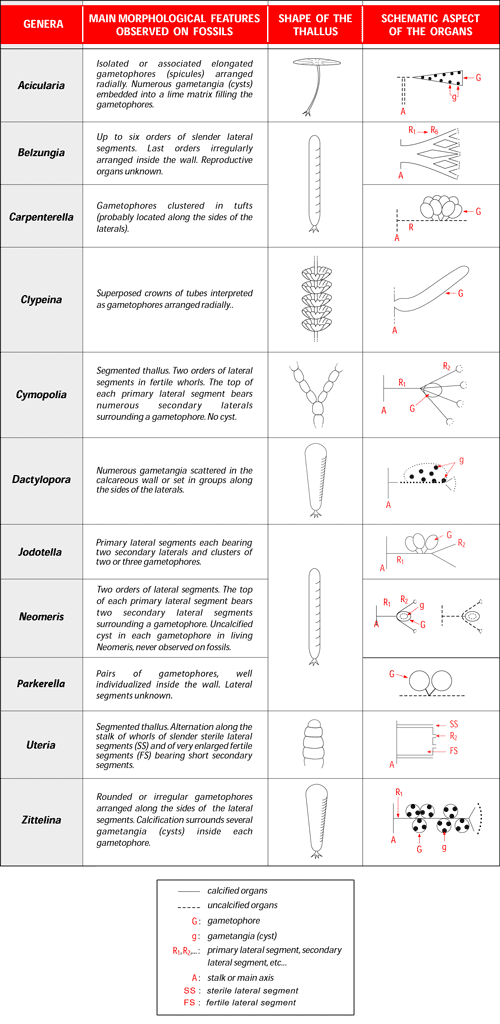
Main morphological features of the species figured in this photo-atlas are already
well-known (Fig. 1 ![]() ). However, almost all the photos are new and some of them show for the first time features that have never been or were incompletely illustrated.
). However, almost all the photos are new and some of them show for the first time features that have never been or were incompletely illustrated.
New information concerns:
- the arrangement of the gametangia, observed in a tangential section of a gametophore in Acicularia eocaenica,
- the internal surface of the main axis showing the location of the proximal ends of the primary laterals in Belzungia borneti,
- the internal surface and the longitudinal section of the tubular calcification of the main axis in Dactylopora cylindracea; the previous photos concerned only the transverse sections ( & , 1954, pl. 5, fig. 2; , 1976, "Dactylopora aff. cylindracea", fig. 3) and the top of the main axis (, 1980, pl. 23, fig. 8),
- the features of the sheath and the morphology of the gametophores in Neomeris defrancei, the arrangement of the units containing the gametophores in Parkerella binodosa; these features were difficult to see on the original illustrations (L. & J. , 1922, pl. 1, figs. 12-18, 53-55) because the photos are too small,
- the longitudinal section of an exceptionally preserved sheath of Neomeris scrobiculata shows the internal surface of the calcification of the main axis and the calcification of several primary laterals; a transverse section of the sheath showing these calcifications had been previously illustrated (, 1987, pl. 8, fig. 6),
- the calcification of the top of the main axis in Uteria encrinella. A comparison with the previously illustrated specimen, regularly rounded at the top (, 1987, pl. 8, fig. 16), suggests that the calcification in this area of the thallus takes various shapes.
Click on the image to enlarge it.
Figure 1: Main morphological features of the genera represented by species in the Ypresian, Thanetian and Bartonian sediments of the Paris basin (drawings: Alain ).
Illustrated species:
- Acicularia eocaenica L. & J. , 1922
- Belzungia borneti L. , 1908
- Carpenterella morelleti , 1980
- Clypeina digitata ( & , 1860) L. & J. , 1913
- Cymopolia zitteli L. & J. , 1913
- Dactylopora cylindracea , 1816
- Jodotella thilense , 1980
- Jodotella veslensis L. & J. , 1913
- Neomeris arenularia ex L. & J. , 1913
- Neomeris craniphora (L. , 1908) , 1927
- Neomeris defrancei (L. & J. , 1922) , 1927
- Neomeris encrinula (, 1822) , 1927 (= Neomeris auversiensis (L. & J. , 1913) , 1927)
- Neomeris herouvalensis ex , 1899
- Neomeris scrobiculata (, 1872) L. & J. , 1913
- Parkerella binodosa L. & J. , 1922
- Uteria encrinella , 1845
- Zittelina dactyloporoides (L. & J. , 1913) , 1987
More information and detailed descriptions of the Cenozoic Dasycladales of the French sedimentary basins are in the main papers of L. & J. (1913, 1917, 1922) and (1978, 1980, 1987, 2009). Features of some Cenozoic species are close to those of Recent species: publications of (1968, 1969), (2006), and (1992) are useful to the interpretation of fossil specimens and to make comparative studies.
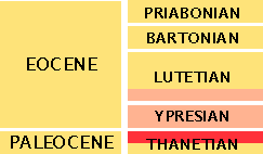
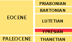
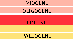
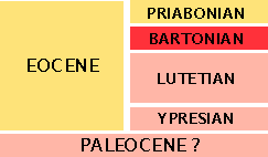
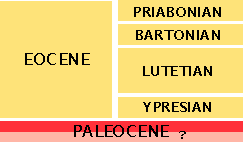
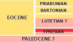
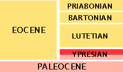
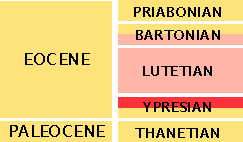
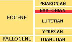
Remark: the stratigraphical range indicated on each slide is the complete stratigraphical range of the species (not just its range in the Paris basin).
|
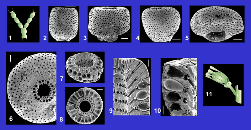
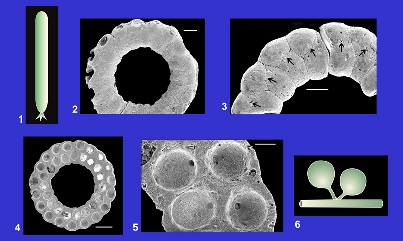
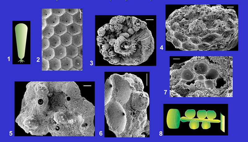
Acicularia eocaenica L. & J. , 1922 |
|
|
Click on the images below to enlarge the photomicrographs |
||
Main references with description and illustrations: L. & J. , 1922, p. 24, pl. 2, figs. 16-20; , 1987, p. 187-193, pl. 14, figs. 1-11. |
||
| Plate 1 | ||
 |
||
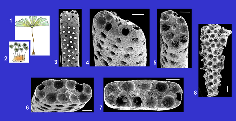
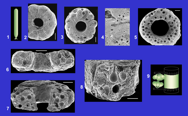
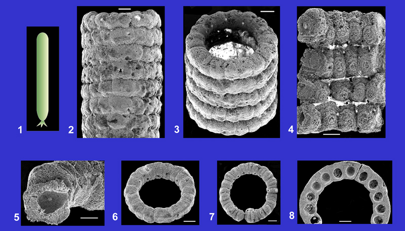
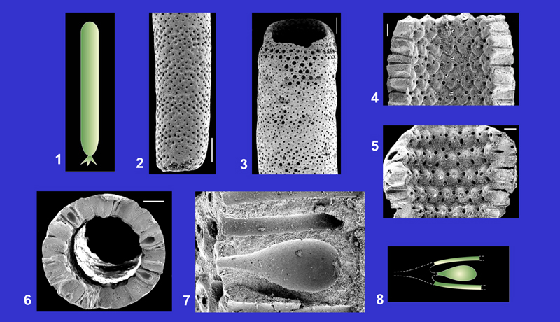
1. Partial hypothetical reconstruction of a living representative. Drawing: Philippe .
2. Hypothetical reconstruction of several living representatives. Drawing: Philippe .
3. External view of a gametophore. Circular openings correspond to the location of the gametangia (cysts). Scale: 200 µm.
4. External view of the distal end of a gametophore. Scale: 100 µm.
5-7. Various shapes of transverse sections and internal aspect of the cavities marking the location of the gametangia. Scales: 100 µm.
8. Longitudinal tangential section of a gametophore showing the arrangement of the gametangia. Scale: 100 µm.
Figs. 3-8: Thanetian, Abbecourt (Oise).
Belzungia borneti L. , 1908 |
 |
|
Click on the images below to enlarge the photomicrographs |
||
|
||
Main references with description and illustrations: L. , 1908, p. 97-99; 1913, p. 38, pl. 3, figs. 38-39; , 1980, p. 22, pl. 25, figs. 1-3; 1987, p. 263, pl. 7, figs. 7-8, 10-11. |
||
| Plate 2 | ||
 |
||
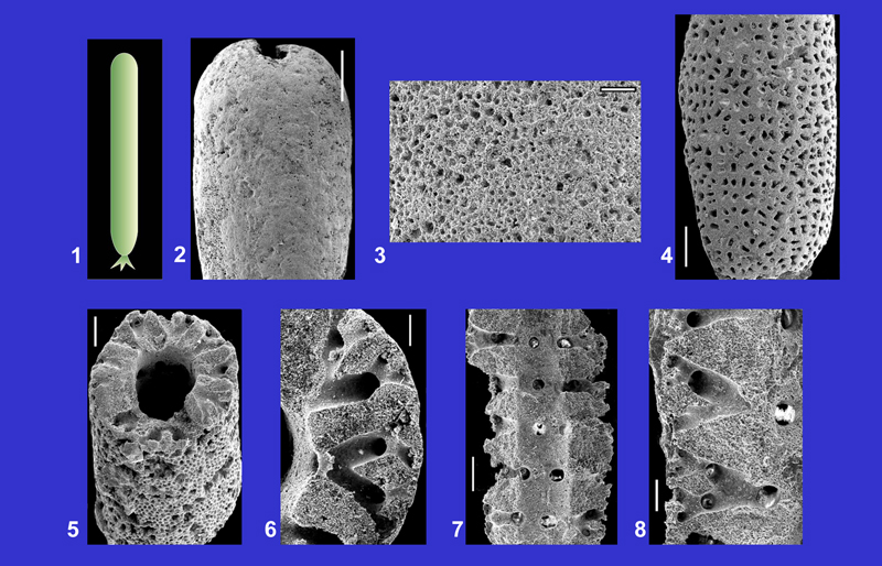
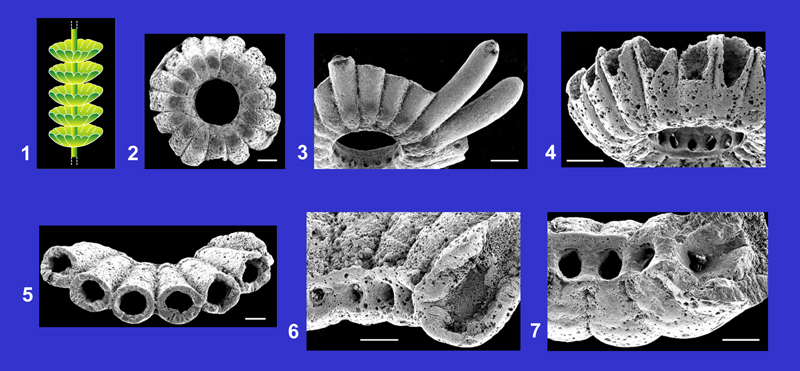
1. Schematic reconstruction of the general shape of the thallus.
2. External view of the sheath. Scale: 500 µm.
3. Detail of the external surface of an irregularly worn specimen. The circular openings with various diameters indicate the location of the fifth and sixth orders of laterals. Scale: 50 µm.
4. Worn specimen showing the arrangement of the third order of laterals. Scale: 200 µm.
5-6. Transverse sections of the sheath traversing several orders of laterals inside the wall. Scales: 100 µm (fig. 5), 50 µm (fig. 6).
7-8. Longitudinal sections of the wall that include several orders of laterals and features of the internal surface of the main axis (fig. 7) showing the regular arrangement of the primary laterals (circular openings). Scales: 100 µm (fig. 7), 50 µm (fig. 8).
Figs. 2-3: Thanetian, Boncourt (Oise); figs. 4-8: Thanetian, Abbecourt (Oise).
Carpenterella morelleti , 1980 |
 |
|
Click on the images below to enlarge the photomicrographs |
||
|
||
Main references with description and illustrations: , 1980, p. 19, pl. 22, figs. 5-11; 1987, pl. 8, figs. 10-11, 14. |
||
| Plate 3 | ||
 |
||
1. Hypothetical reconstruction of a living representative.
2-3. Rings composed of several associated units, each containing a cluster of gametophores. Scales: 100 µm.
4. Groups of small openings corresponding to the location of the peduncles of the gametophores and longitudinal section of a gametophore. Scale: 20 µm.
5. Transverse section of a ring showing the transverse and oblique sections of the numerous gametophores. Scale: 100 µm.
6-8. Longitudinal sections showing the pedunculate gametophores inside the wall. Scales: 100 µm (figs. 6-7) and 50 µm (fig. 8).
9. Reconstruction of two clusters of gametophores located along the laterals. Drawing: Robert .
Figs. 2-8: Upper Ypresian (Cuisian), Hérouval (Oise).
Clypeina digitata ( & , 1860) L. & J. , 1913 |
 |
|
Click on the images below to enlarge the photomicrographs |
||
|
||
Main references with description and illustrations: L. & J. , 1922, p. 27, pl. 2, figs. 37-44 (Cl. pezanti, synonymous); , 1987, p. 237-243, pl. 3, fig. 13, pl. 11, figs. 11-12, pl. 28, figs. 1-10. |
||
| Plate 4 | ||
 |
||
1. Hypothetical reconstruction of a living representative. Drawing: Alain .
2. Lower side of a whorl of gametophores. Scale: 200 µm.
3. Lower side of a whorl with two complete gametophores. Scale: 200 µm.
4. Lower side of a whorl of gametophores. A row of openings located on the internal surface of the axial cavity marks the locations of the proximal ends of the gametophores. Scale: 200 µm.
5. Upper side and transverse sections of several gametophores. Scale: 100 µm.
6-7. Proximal ends of several gametophores (circular openings) and a longitudinal section of the lower end of a gametophore (fig. 6). Scales: 100 µm.
Figs. 2, 4-7: Bartonian (Auversian), Ronquerolles (Val-d'Oise); fig. 3: Lutetian, Montjavoult (Oise).
Cymopolia zitteli L. & J. , 1913 |
 |
|
Click on the images below to enlarge the photomicrographs |
||
Main references with description and illustrations: L. & J. , 1913, p. 11-12, pl. 1, figs. 13-24; , 1980, p. 18, pl. 2, figs. 1-11, pl. 3, figs. 1-5; 1987, p. 289-290, pl. 2, fig. 3, pl. 11, figs. 7-10. |
||
| Plate 5 | ||
 |
||
1. Schematic reconstruction of the general shape of the thallus.
2-5. External views showing the great morphological variability of the articles. The small circular openings mark the location of the secondary laterals. Scales: 500 µm (fig. 2), 200 µm (figs. 3-5).
6. Upper end of an article. Scale: 100 µm.
7. Worn specimen showing the transverse sections of the gametophores (larger openings) surrounded by the secondary laterals (small openings). Scale: 100 µm.
8. Transverse section crossing a circular row of gametophores and several secondary laterals. Scale: 200 µm.
9-10. Longitudinal sections of the upper ends of two articles showing the two orders of laterals and the gametophores. Scales: 100 µm.
11. Reconstruction of a primary lateral segment bearing several secondary laterals and a gametophore. Drawing: Robert .
Figs. 2-3, 5, 7: Bartonian (Auversian), Valmondois (Val-d'Oise); figs. 4, 6, 8-10: Bartonian (Auversian), Le Fayel (Oise).
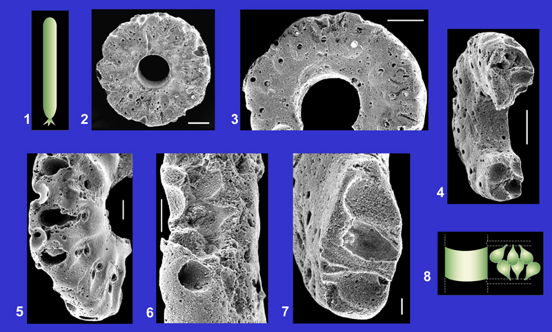
Dactylopora cylindracea , 1816 |
 |
|
Click on the images below to enlarge the photomicrographs |
||
|
||
Main references with description and illustrations: L. & J. , 1913, p. 26-27, pl. 3, figs. 1-4; & , 1954, p. 325-336, pl. 5, figs. 1-4, pl. 6, figs. 5-8, pl. 7, fig. 9; & , 1976, p. 15, pl. 3, figs. 1-2, pl. 4, figs. 1-4; , 1980, p. 21, pl. 23, figs. 1-8. |
||
| Plate 6 | ||
 |
||
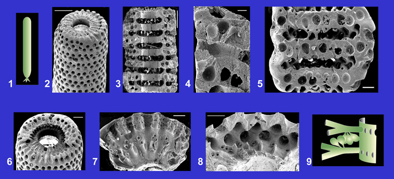
1-2. External views of the sheath. Scales: 1 mm.
3. Longitudinal section and internal surface of the sheath showing the arrangement of the primary laterals (circular openings). Scale: 1 mm.
4. Internal features of the axial cavity with the exceptional preservation of the calcification of the main axis. Scale: 500 µm.
5. Detail of the wall containing groups of gametangia (cysts) inside the partially differentiated gametophores. Scale: 200 µm.
6. Internal features of a gametophore containing several gametangia. Scale: 100 µm.
7. Transverse section of the sheath (the calcification of the main axis is not preserved in this specimen). Scale: 1 mm.
8. Reconstruction of a primary lateral segment bearing three gametophores with several gametangia inside. Drawing: Robert .
Fig. 1: Bartonian (Auversian), Auvers-sur-Oise (Val-d'Oise); figs. 2-7: Bartonian (Auversian), Le Fayel (Oise).
Jodotella thilense , 1980 |

|
|
Click on the images below to enlarge the photomicrographs |
||
|
||
Main references with description and illustrations: , 1980, p. 19, pl. 22, figs. 1-4; 1987, p. 293, pl. 7, figs. 14, 17-18. |
||
| Plate 7 | ||
 |
||
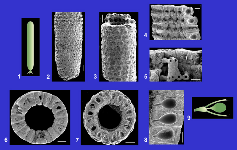
1. Schematic reconstruction of the general shape of the thallus.
2-3. Rings corresponding to the calcification of the gametophores. The circular openings at the surface of the rings show the location of the peduncles of the gametophores. Laterals are not calcified. Scales: 100 µm.
4. Longitudinal section of a ring showing the location of some gametophores (ovoid cavities). Scale: 100 µm.
5. Worn specimen showing the arrangement, more or less in lineations, of the proximal ends of the peduncles (circular openings) and the longitudinal section of a pedunculate gametophore. Scale: 50 µm.
6-7. Longitudinal sections showing several pedunculate gametophores inside the wall. Scales: 50 µm (fig. 6), 20 µm (fig. 7).
8. Reconstruction of two rows of gametophores along the laterals. Drawing: Robert .
Figs. 2-7: Thanetian, Thil (Marne).
Jodotella veslensis L. & J. , 1913 |
 |
|
Click on the images below to enlarge the photomicrographs |
||
|
||
Main references with description and illustrations: L. & J. , 1913, p. 29-30, pl. 3, fig. 12; 1922, p. 16, pl. 1, figs. 49-52; , 1980, p. 18-19, pl. 21, figs. 1-2; et alii, 1985, p. 11-14, pl. 7, figs. 1-10, pl. 8, figs. 1-8. |
||
| Plate 8 | ||
 |
||
1. Schematic reconstruction of the general shape of the thallus.
2. External view of the sheath. The numerous circular openings show the location of the upper ends of the secondary laterals. Scale: 400 µm.
3. Longitudinal section and features of the internal surface of the sheath. Scale: 400 µm.
4. Detail of a longitudinal section showing the pedunculate gametophores at the upper part of a partially preserved primary lateral. Scale: 50 µm.
5. Tangential section of the wall showing transverse, oblique and longitudinal sections of the gametophores. The deep furrows correspond to the location of the partially calcified primary laterals. Scale: 100 µm.
6. Transverse section of the sheath across the location of the laterals (lower part of the photo) and the gametophores (upper part). Scale: 200 µm.
7. Detail of a transverse section across the partially calcified laterals. The groups of two or three circular openings indicate the location of the proximal ends of the pedunculate gametophores. Scale: 100 µm.
8. The large circular openings correspond to the calcification surrounding the secondary laterals. Scale: 100 µm.
9. Reconstruction of two primary laterals, each bearing groups of two or three gametophores and two secondary laterals. Drawing: Robert .
Figs. 2, 6: Thanetian, Jonchery-sur-Vesle (Marne); figs. 3, 7: Thanetian, Abbecourt (Oise); figs. 4-5, 8: Thanetian, Thil (Marne).
Neomeris arenularia ex L. & J. , 1913 |
 |
|
Click on the images below to enlarge the photomicrographs |
||
|
|
||
Main references with description and illustrations: L. & J. , 1913, p. 22-23, pl. 2, figs. 10-14; , 1980, p. 15, pl. 9, figs. 1-11, pl. 10, figs. 11-12, pl. 11, figs. 13-14, pl. 12, fig. 7; 1987, p. 315-316, pl. 5, figs. 14-15, pl. 11, figs. 1-3. |
||
| Plate 9 | ||
 |
||
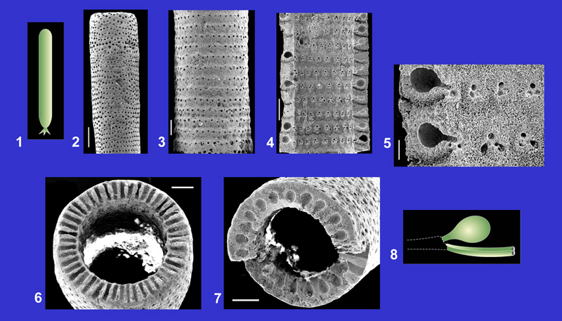
1. Schematic reconstruction of the general shape of the thallus.
2-3. External views of the cylindrical sheath. Single or pairs of openings correspond to the location of the secondary laterals. Scales: 500 µm (fig. 2), 400 µm (fig. 3).
4-5. Internal surfaces of the sheath showing the various shapes of the units that indicate calcification of the gametophores. Primary laterals are not calcified on these specimens. Scales: 200 µm (fig. 4), 100 µm (fig. 5).
6. Internal surface and longitudinal section of the sheath showing numerous gametophores. The calcification of the distal ends of the primary laterals is preserved on the internal surface of the axial cavity. Scale: 200 µm.
7. Transverse section of the sheath across several gametophores. Scale: 200 µm.
8. Reconstruction of the calcified organs: distal end of a primary lateral bearing two secondary laterals and a gametophore. Drawing: Alain .
Figs. 2-5, 7: Bartonian (Auversian), Le Fayel (Oise); fig. 6: "Bartonien", Montagny (Oise), coll. .
 |
||
Click on the images below to enlarge the photomicrographs |
||
Main references with description and illustrations: L. , 1908, p. 96-97, figs. 1a-1d; L. & J. , 1913, p. 15-16, pl. 1, figs. 43-47; , 1970, p. 361-368, pl. 4, figs. 1-14, pl. 5, figs. 1-7; , 1980, p. 15-16, pl. 13, figs. 1-9, pl. 18, figs. 1-2, pl. 19, figs. 1-2, pl. 20, figs. 1-2; 1987, p. 327-328, pl. 5, figs. 1, 5, 6, pl. 7, figs. 1-6. |
||
| Plate 10 | ||
 |
||
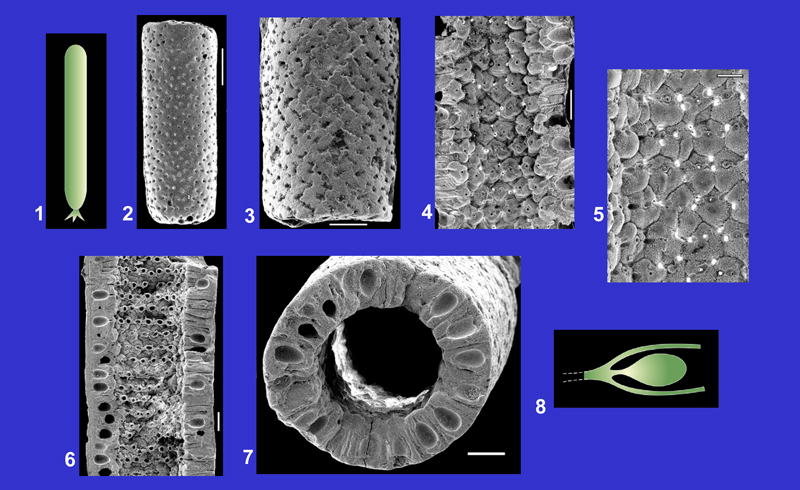
1. Schematic reconstruction of the general shape of the thallus.
2-3. External views of the sheath. The openings indicate the location of the secondary laterals. Scales: 500 µm (fig. 2) and 200 µm (fig. 3).
4. Longitudinal section and features of the internal surface of the axial cavity. Scale: 200 µm.
5. Detail of the longitudinal section of the sheath showing two gametophores and the secondary laterals below the gametophores. Primary laterals are not calcified. Scale: 50 µm.
6. Transverse section across a row of secondary laterals. Scale: 200 µm.
7. Transverse section across two superposed circular rows of gametophores. Scale: 200 µm.
8. Reconstruction of the calcified organs: a gametophore and two secondary laterals. Drawing: Robert .
Figs. 2-3, 6-7: Thanetian, Boncourt (Oise); figs. 4-5: Thanetian, Abbecourt (Oise).
 |
||
Click on the images below to enlarge the photomicrographs |
||
|
||
Main references with description and illustrations: L. & J. , 1922, p. 11, pl. 1, figs. 12-18. |
||
| Plate 11 | ||
 |
||
1. Schematic reconstruction of the general shape of the thallus.
2. External view of the sheath. Each ring corresponds to the calcification of a circular row of gametophores. Scale: 100 µm.
3. External view and transverse section of the sheath. Scale: 100 µm.
4. Internal surface of the axial cavity and longitudinal section of the sheath. Each unit is the calcification of a gametophore. The primary laterals are not calcified. The canals inside the wall (left side of the photo) show the location of the partially preserved secondary laterals. Scale: 100 µm.
5. Detail of a longitudinal section showing a gametophore inside the wall. Scale: 50 µm.
6-7. Isolated rings. The small circular openings located on the internal side of the ring correspond to the proximal ends of the gametophores. Scales: 100 µm.
8. Transverse section of the sheath crossing a circular row of gametophores. Scale: 100 µm.
Figs. 2-8: Thanetian, Abbecourt (Oise).
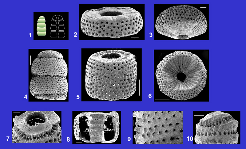
Neomeris encrinula (, 1822) , 1927 |
 |
|
Click on the images below to enlarge the photomicrographs |
||
Remark: Neomeris auversiensis (L. & J. , 1913) , 1927 is considered junior synonym of N. encrinula ( & , 2011). |
||
Main references with description and illustrations: L. & J. , 1913, p. 16-17, pl. 1, figs. 48-50; 1939, p. 30-33, pl. 4, figs. 7-20; , 1980, p. 17, pl. 16, figs. 10-12, pl. 17, figs. 1-11, pl. 18, figs. 7-8, 12-14, pl. 19, figs. 11-13, pl. 20, fig. 6-7; 1987, p. 317, 328-329, pl. 11, figs. 4-6, pl. 35, figs. 1-5; & , 2011, pl. 3, figs. 1-8. |
||
| Plate 12 | ||
 |
||
1. Schematic reconstruction of the general shape of the thallus.
2. External view of the sheath. The surface is characterized primarily by its longitudinal ridges. The circular rows of small openings correspond to the whorls of secondary laterals. Scale: 300 µm.
3. Transverse section of the sheath and detail of the external surface showing the regular rows of secondary laterals. Scale: 200 µm.
4. Detail of the axial cavity showing the well individualized calcification of the main axis. Scale: 100 µm.
5. Tangential section of the wall showing the transverse sections of several gametophores (large circular openings). Scale: 100 µm.
6. Transverse section across a circular row of secondary laterals. Scale: 100 µm.
7. Features of the axial cavity and longitudinal section of the wall across several gametophores. The calcification of the main axis is not preserved in this specimen. Scale: 100 µm.
8. Longitudinal section of the wall showing a gametophore and two secondary laterals. Scale: 50 µm.
9. Reconstruction of a gametophore and two secondary laterals. Primary laterals are unknown. Drawing: Alain .
Figs. 2, 5, 7: Bartonian (Auversian), Val-d'Oise; figs. 3-4, 6, 8: Bartonian (Auversian), Mériel (Val-d'Oise).
Neomeris herouvalensis ex , 1899 |
 |
|
Click on the images below to enlarge the photomicrographs |
||
Main references with description and illustrations: L. & J. , 1913, p. 23, pl. 2, figs. 21-23; , 1980, p. 13, pl. 5, figs. 1-8, pl. 10, figs. 1-2, pl. 11, figs. 1-2, pl. 12, fig. 1; 1987, p. 334-335, pl. 2, fig. 5, pl. 5, fig. 7, pl. 8, figs. 7-9. |
||
| Plate 13 | ||
 |
||
1. Schematic reconstruction of the general shape of the thallus.
2. External view of the sheath. The circular openings show the location of the secondary laterals. Scale: 500 µm.
3. Worn specimen showing the transverse sections of the gametophores (large circular openings at the top of the sheath) and the transverse sections of the secondary laterals (small circular openings). Scale: 300 µm.
4-5. Different features of the internal surface of the sheath. Each unit indicates the calcification of a gametophore bounded by two secondary laterals. The main axis, the primary laterals and the lower part of the secondary laterals are never calcified. Scales: 100 µm.
6. Transverse section of the sheath across several gametophores and secondary laterals. Scale: 200 µm.
7. Detail of the wall showing a gametophore and several secondary laterals. Scale: 30 µm.
8. Reconstruction of a gametophore together with two partially preserved secondary laterals. Drawing: Alain .
Figs. 2-7: Upper Ypresian (Cuisian), Hérouval (Oise).
Neomeris scrobiculata (, 1872) L. & J. , 1913 |
 |
|
Click on the images below to enlarge the photomicrographs |
||
Main references with description and illustrations: L. & J. , 1913, p. 21-22, pl. 2, fig. 20; 1922, p. 14, pl. 1, fig. 38; , 1980, p. 14, pl. 4, figs. 1-11, pl. 10, figs. 3-4, pl. 11, figs. 3-4, pl. 12, fig. 2; 1987, p. 353-354, pl. 5, figs. 8-9, pl. 8, figs. 1-6. |
||
| Plate 14 | ||
 |
||
1. Schematic reconstruction of the general shape of the thallus.
2-3. External views showing the typical feature of the wall: single or pairs of rounded swellings surrounding the location of the secondary laterals (small circular openings). Scales: 200 µm.
4. Internal surface of the cylindrical sheath showing the calcification of four superposed rows of gametophores and longitudinal section of the wall crossing several gametophores. Scale: 100 µm.
5. Calcification of the main axis preserved exceptionally inside the axial cavity of the sheath. Several primary laterals are also calcified. Scale: 100 µm.
6. Transverse section. Each unit corresponds to the calcification of a gametophore. Scale: 200 µm.
7. Transverse section across a circular row of gametophores. Scale: 200 µm.
8. Longitudinal section of three pedunculate gametophores. Scale: 100 µm.
9. Reconstruction of a primary lateral bearing two secondary laterals and a gametophore. Drawing: Alain .
Figs. 2, 4, 6-8: Upper Ypresian (Cuisian), Pierrefonds (Oise); figs. 3, 5: Upper Ypresian (Cuisian), Cuise-la-Motte (Oise).
Parkerella binodosa L. & J. , 1922 |
 |
|
Click on the images below to enlarge the photomicrographs |
||
|
Main references with description and illustrations: L. & J. , 1922, p. 16, pl. 1, figs. 53-55; , 1978, p. 219-224, text.-fig. 22; 1987, p. 359-363, tabl. 33. |
||
| Plate 15 | ||
 |
||
1. Schematic hypothetical reconstruction of the general shape of the thallus.
2. External view of a ring corresponding to the calcification of the gametophores. Laterals are not calcified (lectotype, specified by , 1987, p. 360; specimen previously figured in L. & J. , 1922, pl. 1, fig. 53). Scale: 100 µm.
3. Fragment of a ring (specimen figured in L. & J. , 1922, pl. 1, fig. 54). Each unit corresponds to the calcification of a gametophore. The arrows indicate the location of the lower ends of the peduncles (pairs of small circular openings) bearing the gametophores. Scale: 100 µm.
4. Transverse section of a ring showing two concentric and alternating rows of gametophores (specimen figured in L. & J. , 1922, pl. 1, fig. 55). Scale: 200 µm.
5. Detail of fig. 4. Sections of several gametophores. The small openings correspond to the transverse sections of the peduncles. Scale: 50 µm.
6. Reconstruction of a pair of gametophores along a lateral (the shape of the lateral is hypothetical). Drawing: Robert .
Figs. 2-5: Thanetian, Abbecourt (Oise), coll. .
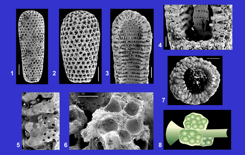
Uteria encrinella , 1845 |
 |
|
Click on the images below to enlarge the photomicrographs |
||
|
|
||
|
Main references with description and illustrations: L. & J. , 1913, p. 39-41, pl. 3, figs. 40-46; 1938, p. 179-181, text.-figs. 1-4; , 1980, p. 22, pl. 25, figs. 8-13; 1987, p. 375, pl. 8, figs. 12-13, 15-16. |
||
| Plate 16 | ||
 |
||
1. Schematic reconstruction of the general shape of the thallus (left) and schematic axial section (right) (L. & J. , 1938, text.-fig. 4, modified).
2, 5. Two specimens showing the morphological variability of the articles. Scales: 200 µm (fig. 2), 500 µm (fig. 5).
3. Side view of an upper article. Scale: 200 µm.
4. Three superposed articles at the upper end of the sheath. Scale: 500 µm.
6. Upper side of the penultimate article. Scale: 500 µm.
7. Internal view of the lower end of an article. The lower part of the photo shows the location of a whorl of laterals. Scale: 200 µm.
8. Longitudinal section of an article showing the calcification of the main axis. Scale: 200 µm.
9. Detail of the internal surface of an article. Scale: 100 µm.
10. Upper end of the calcification of the main axis. Scale: 100 µm.
Figs. 2-3, 6, 10: Upper Ypresian (Cuisian), Hérouval (Oise); figs. 4-5, 7-9: Upper Ypresian (Cuisian), Liancourt-Saint-Pierre (Oise).
 |
||
Click on the images below to enlarge the photomicrographs |
||
|
||
Main references with description and illustrations: L. & J. , 1913, p. 28-29, text.-figs. 14-16; , 1987, p. 376, pl. 1, fig. 13, pl. 2, fig. 9, pl. 38, figs. 1-12. |
||
| Plate 17 | ||
 |
||
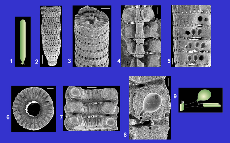
1. Schematic reconstruction of the general shape of the thallus.
2. Detail of the external surface of the sheath. The cortical layer is composed of alveoli that indicate the enlarged distal ends of the laterals. Scale: 200 µm.
3. Transverse section across the calcification of the main axis and several laterals. The gametophores are sited along the laterals and contain numerous gametangia (small rounded cavities). Scale: 200 µm.
4. Transverse section of a specimen containing gametophores very irregular in shape. Scale: 100 µm.
5. Detail of the wall showing several gametophores enclosing the laterals (circular openings). Scale: 100 µm.
6. Detail of the wall showing a gametophore near the alveolate cortical layer. Scale: 100 µm.
7. Internal feature of a gametophore showing the location of several gametangia. Scale: 20 µm.
8. Reconstruction of a primary lateral bearing several gametophores. Drawing: Robert , modified.
Figs. 2-3, 5-6: Bartonian (Auversian), Baron (Oise); figs. 4, 7: Bartonian (Auversian), Echampeu (Aisne).
We thank Alain and Florentin for creating the plates and several drawings, Alain and Nicolas (Scanning Electron Microscopy and Microanalysis Center, University of Nantes) for their assistance with scanning electron microscopy, Robert and Philippe for the reconstructions of the species.
F. & P. (1976).- Osservazioni sul genere Dactylopora 1816 (Alghe versi Dasicladacee).- Bollettino della Società dei Naturalisti in Napoli, vol. LXXXV, p. 179-203 (25 p.).
S. (2006).- Photo-Atlas of living Dasycladales.- Carnets de Géologie - Notebooks on Geology, Brest, Book / Livre 2006/02 (CG2006_B02), 348 p.
S. & M.J. (1992).- Dasycladales. An illustrated monograph of a fascinating algal order.- Georg Thieme Verlag, Stuttgart, 247 p.
J. (1976).- Genus Dactylopora , Digitella & und Broeckella & (Dasycladaceae, Algae) in kalken des Palaozans der Westkarpaten.- Geologicky Zbornik - Geologica carpathica, Bratislava, vol. 27, n° 2, p. 247-272.
R. (1970).- Niveau à algues dans le Sparnacien de la région de Plagne (Petites-Pyrénées, Haute-Garonne) et observations sur le genre Neomeris 1816.- Bulletin du Centre de Recherches Pau - SNPA, vol. 4, n° 2, p. 353-379.
I., F. & R. (1985).- Paleocene Dasycladalean algae from Orosei (Eastern Sardinia).- Memorie degli Istituti di Geologia e Mineralogia dell'Università di Padova, vol. XXXVIII, 77 p.
P. (1978).- Les Dasycladacées du Paléocène supérieur et de l'Éocène du Bassin de Paris.- Thèse Doctorat en Sciences de la Terre, Bordeaux I, 403 p.
P. (1980).- Les Dasycladacées du Paléocène supérieur et de l'Éocène du Bassin de Paris.- Mémoires de la Société géologique de France, Paris, (N.S.), t. LIX, n° 138, 40 p., 25 pls. (I-XXV).
P. (1987).- Les Chlorophycées calcaires du Paléogène d'Europe Nord-Occidentale (Bassin de Paris, Bretagne, Cotentin, Bassin de Mons).- Thèse Doctorat d'État, Nantes, 500 p.
P. (2009).- Cenozoic Dasycladales. A photo-atlas of Lutetian species from French Cenozoic basins.- Carnets de Géologie - Notebooks on Geology, Brest, Special Publication 2009/01 (CG2009_SP01); ISBN 978-2-916733-03-6, Book / Livre 2009/01 (CG2009_B01), 180 p.
P. & J. (2011).- New species and data on Lutetian Dasycladales (calcareous algae) of Cotentin (Normandy, France).- Revista Española de Micropaleontología, Madrid, vol. 43, n° 1-2, p. 141-155.
J.B.P.A. (1816).- Histoire naturelle des animaux sans vertèbres. Tome IIème. Histoire des Polypes.- Verdière (Libraire), Paris, 568 p.
G. & T. (1954).- Microstructure d'une algue calcaire: Dactylopora (Dasycladacée tertiaire). Regards sur les organismes aragonitiques et sur leur fossilisation.- Revue générale de Botanique, Paris, t. 61, p. 325-336.
H. (1845).- Iconographie zoophytologique. Description par localités et terrains des Polypiers fossiles de France et pays environnants.- P. Bertrand (Éditeur), Paris, 348 p.
L. (1908).- Deux algues siphonées verticillées du Thanétien de Boncourt (Oise).- Bulletin de la Société géologique de France, Paris, (4e Série), t. VIII, p. 96-99.
L. & J. (1913).- Les Dasycladacées du Tertiaire parisien.- Mémoires de la Société géologique de France, Paris, t. XXI, fasc. 1, n° 47, 43 p., 3 pls. (I-III).
L. & J. (1917).- Les Dasycladacées tertiaires de Bretagne et du Cotentin.- Bulletin de la Société géologique de France, Paris, (4e Série), t. XVII, fasc. 6-7, p. 362-372, 1 pl. (XIV).
L. & J. (1922).- Nouvelle contribution à l'étude des Dasycladacées tertiaires.- Mémoires de la Société géologique de France, Paris, t. XXV, fasc. 2, n° 58, 35 p., 2 pls. (IX-X).
L. & J. (1939).- Tertiary Siphoneous Algae in the W.K. Collection with descriptions of some Eocene Siphoneae from England.- British Museum (Natural History), London, 55 p., 6 pls. (I-VI).
M. (sic) = L. & J. (1938).- Les algues siphonées calcaires. In: L. & G., Révision des collections H. .- Bulletin du Muséum national d'Histoire naturelle, Paris, (2e Série), t. X, n° 2, p. 178-183.
J. (1927).- 1. Abteilung: Thallophyta. In: M., Handbuch der Paläeobotanik.- Oldenbourg, München u. Berlin, Band I, p. 31-112.
G. (1899).- Ueber fossile Dasycladaceen vom Cerro Escamela, Mexico.- Botanische Zeitung, Leipzig, Heft VIII, p. 137-154.
G. (1968).- Contribution à l'étude des Dasycladales. 1. Morphogenèse.- Nova Hedwigia, Lehre, Band XVI, p. 21-82.
G. (1969).- Contribution à l'étude des Dasycladales. 2. Cytologie et reproduction. 3. Révision systématique.- Nova Hedwigia, Lehre, Band XVII, p. 551-644.
P. & B. (2011).- Cenozoic Dasycladales. A photo-atlas of Thanetian, Ypresian and Bartonian species from the Paris basin.- Carnets de Géologie - Notebooks on Geology, Brest, Special Paper 2011/01 (CG2011_SP01), 44 p., 1 fig., 17 pls.