 |
|
|||
|
Genus Novocrania Lee et Brunton, 2001 |
|
[Type species = Patella anomala Müller, 1776; ICZN plenary powers, 1988, opinion 1468] |
| Dorsal valve convex to conical; beak subcentral to posterocentral, smooth, finely pustulose or rarely finely costellate; posterior margin commonly straight; recent species with dendroid shell punctation; dorsal posterior adductor scars large, rounded, thickened, widely separated; anterior scars commonly crescentic, raised above valve floor; weak myophragm bisects muscle field; encrusting; ventral valve uncalcified in recent species, otherwise sometimes thin; ventral posterior adductor scars large, anterior scars united medially; marginal mantle setae observed in recent forms; valve margins variably thickened, with limbus or faint submarginal rim. |
|
Paleogene (Eocene) - Present
Diagnosis from Lee & Brunton (1986), Lee (1987), and
|
| Extant Species of Novocrania |
|
syn : N. turbinata (Poli, 1795) | Diagnosis Diagnosis Diagnosis Diagnosis Diagnosis |
|
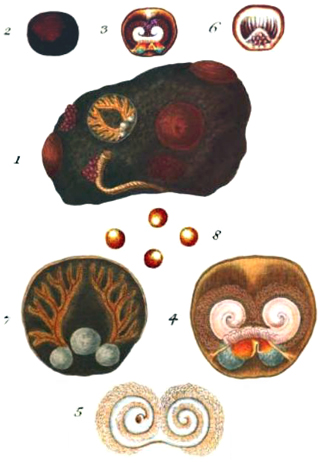
Novocrania anomala (Müller, 1776) - type species Type locality: Hår-Krøllen (Denmark) Patella anomala Müller, 1776, p. 237 Diagnosis of Robinson (2017) Dorsal valve exterior smooth to hummocky with concentric growth lamellae, dorsal valve interior with adductor muscle scars flush with valve surface or slightly raised, small anterior muscles commonly attached to a tiny anterior septum, ventral valve sometimes organic, commonly partly calcitic/partly organic or completely calcitic, ventral valve punctate (when calcitic), rostellum organic, ventral posterior adductor muscle scars organic, in dead shells ventral muscle scars and rostellum latent. |

Original Diagnosis Müller (1776) 2870. Patella anomala, testa rudi, sulca, orbiculari, vertice submarginali. |
||
|
|
|||
|
Novocrania turbinata (Poli, 1795) Type locality: Sicily (Italy) Patella kermes da Costa et Humphey, in da Costa, 1770 Crania suessi Reeve, 1862 Crania japonica Adams, 1863 Diagnosis of Robinson (2017) Dorsal valve exterior commonly minutely spinose or pustulose, dorsal valve interior with anterior adductor muscles and support structure attached to raised pedestals, small anterior muscles attached to median process, ventral valve has radial canals, is mostly impunctate, the calcitic rostellum has a slim spike, the ventral anterior adductor muscle scars are calcitic or uncommonly latent. Robinson (2017, p. 517) states: "The dorsal valve is sub-conical, the exterior has a hummocky surface with an ornament of concentric growth rings [as in N. anomala?] and, in many populations, specimens may have minute pustules or spines". |
 Robinson (2017) establishing this species as valid did not studied specimens from the type-locality, nor from the Mediterranean bathyal. This species may occur together with N. anomala, but even with large material available in many Mediterranean areas, no extensive study has been performed on these Novocrania: consequently, their geographic and bathymetric distribution cannot be established. More in Emig (2014)... |
||
|
|||
|
|
|||
|
Novocrania lecointei (Joubin, 1901) Type locality: “N° 578b. - Faubert VII – Lat.70°23'S., Long. 82°47'O - 8 Octobre 1898 – Profondeur : 500 m environ.” ? Crania pourtalesii Dall, 1890, p. 232 (not Crania anomala var. pourtalesii Dall, 1871, p. 35) Diagnosis of Robinson (2017) Dorsal valve varies from smooth to hummocky, with weak to strong concentric growth rings/lamellae, to spinose with tubular hollow spines, usually broken short, dorsal valve exterior white to grey, ventral valve organic with organic rostellum. Diagnosis should be completed at least by reproduction characters (see in front). |
Original description by Joubin (1901) - in facsimile
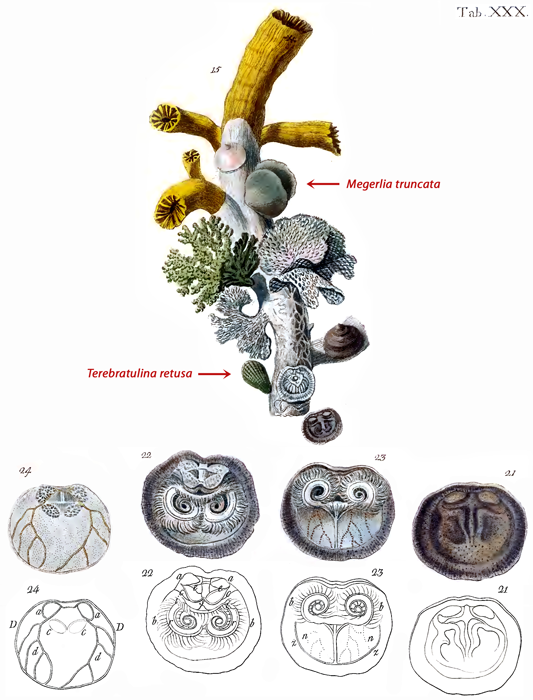
Genre CRANIA. Crania Lecointei n. sp. PI. II, fig. i3, 14 et 15. N° 578 b. — Faubert VII. — Lat. 70°23' S., Long. 82°47' O. — 8 Octobre 1898. — Profondeur : 5oo m environ. Couleur de l'animal vivant : Ochraceus à Bruneus clair (cf. Chromotaxia de Saccardo). Cette pêche a rapporté deux échantillons de cette Crania fixés chacun sur un petit galet ; l'un est intact, l'autre brisé. près sous la crête qui, partant du sommet pédonculaire, va aboutir au-dessus des cruras. Cette disposition se voit nettement dans la figure 4. Il en résulte que la région terminale de la valve est découpée par ces deux cloisons en trois pyramides, le pédoncule occupant celle du milieu. Je ne puis presque rien dire de l'animal n'ayant pu en extraire que quelques fragments. Les glandes génitales, très peu ramifiées, et contenant des œufs murs, n'étaient développées que dans la paroi de la cavité viscérale, et nullement dans ses dépendances palléales. Je n'ai pu voir la forme des sinus palléaux. L'un des bras avait 13mm de long, il ne m'a pas paru différer de ce que l'on sait de ces organes chez R. psittacea. Le cul-de-sac postérieur du tube digestif est droit et non renflé en vésicule. Les cœcums de la glande hépatique ont une grande longueur. |
||
|
|||
|
|
|||
|
Novocrania huttoni (Thomson, 1916) Type locality: "Whangaroa, Cook Strait" - [New Zealand]. Crania huttoni Thomson, 1915, p. 41-42 Diagnosis of Robinson (2017) Dorsal valve exterior may be smooth or have fine radial costae, rarely developed into minute spines, dorsal valve exterior often mottled, brown-reddish brown and white, ventral valve organic with non-crystalline calcareous rostellum. |
There is in the Dominion Museum a tablet holding three valves from an unknown locality which purport to be the specimens upon which Hutton based the above brief description. All three valves, however, appear to be dorsal valves, and the apparent absence of radiating lines on two of them is due in one case to attrition on the sea-bottom and in the other to an encrusting organism. There is also in the Museum another tablet of seven valves- which are those mentioned by Hamilton as '' Trail ; Whangaroa, Cook Strait."' Of these seven, the two smallest are the young of Anomia sp., but the other five are dorsal valves of the same species of Crania as Hutton's specimens. In view of the locality record attaching to the latter specimens, the best preserved of these is chosen as the holotype. (Thomson, 1916, p. 41). | ||
|
|
|||
|
Novocrania philippinensis (Dall, 1920) Type locality: "Between Masbate and Leyte Islands, Philippines, in 114 fathoms, green mud, at Bureau of Fisheries station 5398." Crania philippinensis Dall, 1920, p. 272 Diagnosis of Robinson (2017) Dorsal valve exterior smooth to hummocky to minutely spinose, anterior adductor muscle scars and support structure scars commonly on raised pedestals, support structure scars commonly placed at lateral tip of anterior adductor muscle scars making the latter scars appear elongated, small anterior muscles attached to median process, ventral valve calcitic and punctate, rostellum calcitic with small anterior muscle scars on rounded tip. |
|||
|
|
|||
