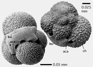
Figure 1: Accessory aperture in bulla of Globigerinita glutinata (). SEM graph from et alii,
1993.
aca: accessory aperture; bu: bulla; ch: ordinary spiral chamber.
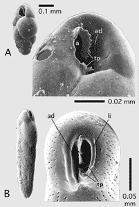
Figure 2: Adapertural depression and toothplate with serrated margin. SEM, from et alii, 1993.
A: Bulimina elongata d'; B: Loxostomina cf. africana ().
a: aperture; ad: adapertural depression; li: lip; tp: toothplate with its serrated margin.
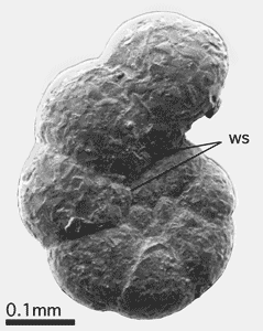
Figure 3: Advolute whorl in agglutinated planispiral shell of Labrospira jeffreysii (). SEM,
from et alii, 1993.
ws: whorl suture.
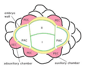
Figure 4: Embryonic and periembryonic chambers in Orbitoides spp. according to 's
1966 concept.
I: protoconch; II: deuteroconch; PAC: principal auxiliary chamber;
PEC: principal epi-auxiliary chamber; AEC: accessory epi-auxiliary chamber. In our view, we prefer to call PAC an auxiliary and AEC an adauxiliary chamber.
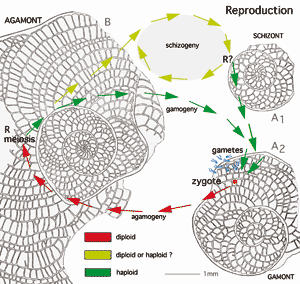
Figure 5: Standard dimorphic or trimorphic reproductive cycle in benthic, medium- to large-sized foraminifera according to ,
1999. Schizogeny, eventually repeated several times, may be widespread in larger foraminifera from oligotrophic habitats. Planktic foraminifera seem to have no dimorphic life cycle. Life cycles are linked in various ways to seasonal cycles. Example: Heterostegina depressa d', equatorial sections, Gulf of Aqaba, Red Sea (from ,
1977).
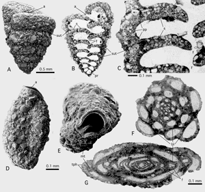
Figure 6: Agglutination in foraminiferan walls; Gulf of Aqaba, Red Sea; Recent.
A-C: Textularia sp. C in et alii, 1993. A: Lateral view showing coarse grains producing a rugged shell surface exept on apertural face. SEM. B: axial thin section of the biserial test showing distribution of agglutinated grains within the shell walls. Light microscope, transparent light. C: Detail from another specimen showing parapore texture of wall and its relation to the agglutinated grains. Transmitted light. D-G: Agglutinella sp., an agglutinating, porcelaneous miliolid. D: lateral view showing coarse agglutination at the shell's suface. E: apertural view showing large aperture with a porcelaneous peristomal rim and a miliolid tooth. SEM.
F-G: Schlumbergerina alveoliniformis (),
thin sections in the - and perpendicular to the - apertural axis, showing early growth stages without apparent agglutination and a very thin basal layer coating the rugged surface of adult chambers of the previous whorl. Transmitted light.
a: aperture; bl: basal layer; f: foramen; mt: miliolid tooth; pp: parapores; pr: proloculus; s: septum; sut: (chamber) suture; tph: trematophore.
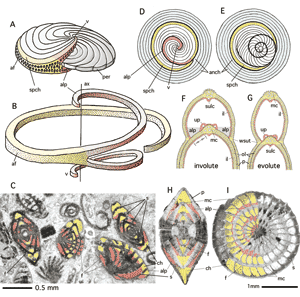
Figure 7: Alar prolongations and involuteness in lamellar foraminifers.
A: planispiral-involute shell with a long apertural face, as in Archaias. Note the apertures in the face of both alar prolongations.
B: shape of single (penultimate) spiral chamber with long alar prolongations forming a vortex. C: Random sections of "Peneroplis" glynnjonesi ,
Lower Oligocene, Iran. Transmitted light. Note foramina in the alar septa. D-E: Transition from planispiral-involute to annular growth. D: external, lateral view of subsequent chambers, showing the ultimate spiral chamber with its alar prolongation and subsequent annular chambers. E: all chambers in equatorial section including the tightly coiled nepiont. F-G: Involuteness and evoluteness in lamellar foraminifers: an accurate definition is whether or not perforate walls (double arrow) cover the next whorl. Axial section, schematic, not to scale. Note the numerous, outer lamellas (green) enveloping the total exposed surface of the previous shell (compare "lamellation"). The distribution of the inner lamella (red) covering the previous whorl hasno significance in the definition of involuteness. H-I: Nummulites incrassatus .
Upper Eocene, Northern Italy. H: not quite centered axial section.
Red: lumen of alar prolongation, yellow: lumen of equatorial chamber. I: oblique section almost perpendicular to shell axis. Note the obliqueness of the intersections of the alar prolongations.
af: apertural face; alp: alar prolongation; anch: annular chambers; ax: shell axis; ch: chamber; f: foramen; il: inner lamella; mc: marginal cord; ol: (numerous) outer lamellas; p: pore; per: periphery (of spiral shell); s: septum; spch: spiral chamber; sulc: sulcus; v: vortex; wsut: whorl suture.
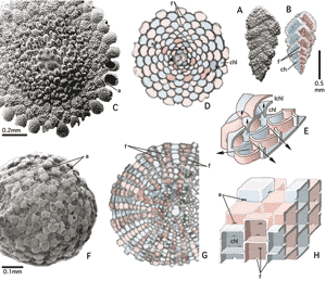
Figure 8: Alternating arrangement of shell compartments in the three dimensions of space. Examples from Gulf of Aqaba, Red Sea; Recent.
A-B: Alternating chamber arrangement in the linear dimension (= biserial arrangement) as in Textularia foliacea et , SEM and X-ray graphs of lateral view. C-D: Alternating chamberlet arrangement in the planar (second) dimension, as in annular-concentric, discoidal shell of Planorbulinella elatensis , SEM graph of lateral view and equatorial section in transmitted light microscopy, coloured. E: Stereograph showing alternating chamberlets of a main chamberlet layer, with main stolon axes (horizontal arrows) and retrovert stolons feeding lateral chamberlet (vertical arrows). Schematic, not to scale. F-G: Alternating chamberlet arrangement in the third dimension producing a chessboard pattern, as in the spherical-concentric, globular shell of Sphaerogypsina globulus (), SEM graph of external view and centered section in transmitted light micrograph, coloured. H: Stereograph showing alternating chamberlets forming chessboard pattern. Schematic, not to scale.
Colours: red and blue: alternating generations of shell compartments; green: nepiontic, early stages including proloculus.
a; aperture; ch: chamber lumen; chl: chamberlet lumen; f: foramen; lchl: lateral chamberlet lumen.
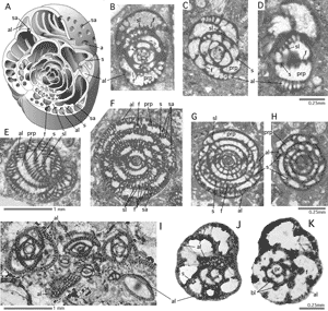
Figure 9: Alveoles.
A-D: model (schematic, not to scale) of the genus Malatyna , subaxial, subequatorial and tangential sections. Igualada, North-Eastern Spain.
Uppermost Middle Eocene. E-F: Globoreticulina iranica , tangential and axial sections. Shiraz, Iran. Middle-Upper Eocene. G-H: Bullalveolina bulloides , axial and subequatorial sections. Peribetics, South-Eastern Spain. Lower Oligocene. I: Austrotrillina striata , sections perpendicular to apertural axis and tangential sections. Kirkuk, Iraq. Oligocene. J-K: Everticyclammina virguliana (), equatorial sections. Mechra Klila, North-Eastern Morocco, Uppermost Jurassic. All sections transmitted light micrographs.
a: (main) aperture; al: alveole; bl: basal layer; f: (main) foramen; pr: proloculus; prp: preseptal passage (in these cases extending over most of the chamber lumen); s: septum; sa: supplementary aperture; sl: septular ridges (incomplete septula).
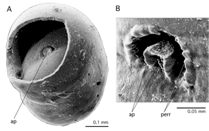
Figure 10: Apertural plate: Apertural feature in Sphaeroidina bulloides d'.
A: dissected specimen showing penultimate foramen, B: detail of aperture. SEM graphs. Recent, Gulf of Aqaba.
ap: apertural plate; perr: peristomal, pustulose rim.
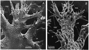
Figure 11: Arborescent shell ornamented by areoli: Homotrema rubra (), arborescent habit, SEM graphs, Recent, Gulf of Aqaba.
A: overview of arborescent shell, B: detail of a single branch, Note the sponge spicules (ssp) glued into the tubular peristomal extension of the aperture to serve as fishing rods that support filiform pseudopodia extending into turbulent water for the capture of food.
a: aperture; ar: areoli.
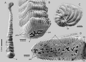
Figure 12: Single and multiple areal
apertures.
A-B: Monalysium acicularis (); C-D: Peneroplis planatus ( et ), both from the Gulf of Aqaba, Red Sea, Recent. SEM graphs. Note the presence of pits in the porcelaneous wall and the umbilical bowl characterizing P. planatus.
a: areal aperture(s); pst: peristomal rims; ub: (delimitation of) umbilical bowl; ws: whorl suture. Note the absence of interiomarginal apertures.
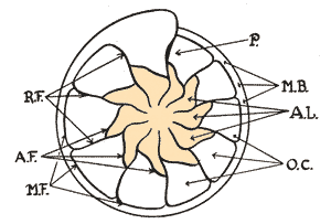
Figure 13: Astral lobes: Original figure from , 1932, p. 414.
AF: astral furrow (= foliar suture); AL (coloured): astral lobes (= folia): MB: marginal band (= keel); MF: marginal furrow (= septal suture); OC: outer chambers; P: pylome (= aperture); RF: radial furrows (= chamber sutures).
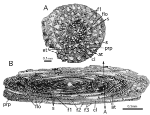
Figure 14: Attics and floors in Alveolinella borneensis , Molukkas, Miocene. Transmitted light micrographs.
A: Transverse section. Position in respect to B: double arrow A. B: axial section.
at: attics; cl: chamberlet; f1: foramen (basal row): f2: foramen (second row); f3: supplementary foramina corresponding to the attics; flo: floors; s: septum.
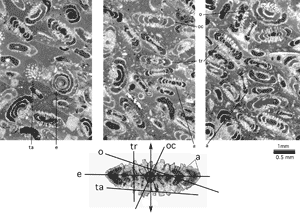
Figure 15: Sections of Involutina liassica (). Middle Lias, from Arzo, Ticino, Southern Switzerland. Transparent light micrographs. Involutina exhibits a lamellar-perforate, planispiral-evolute, dimorphic shell with a spherical megalosphere followed by a tubular chamber cavity without septa. Lateral walls ornamented by protuberant piles between coarse pores. Double arrow: shell axis.
a: axial section; e: equatorial section; o: oblique section; oc: oblique-centered section; ta: tangential section; tr: transverse section.
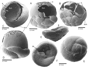
Figure 16: Balloon and float chambers in pseudoplanktic forms. All specimens from the Gulf of Aqaba, Red Sea, Recent. SEM graphs.
A-B: Cymbaloporetta sp. Lateral views of dissected specimens. Note tubular infolds of the float chamber canalizing the hatching from the umbilical cavity to the preseptal space under the apertures of the balloon chamber (arrows). C-D: Tetromphalus bulloides (d'). Lateral views of dissected and intact specimens in their pseudoplanktic stage. E-G: T. bulloides in its benthic stage, showing open umbilicus, lateral, umbilical (ventral) and spiral (dorsal) views.
a: aperture; bch: balloon chamber; flch: float chamber; fol: folium; u: umbilicus.
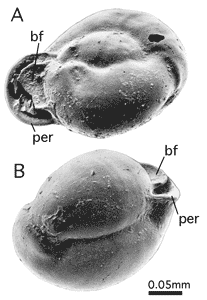
Figure 17: Basal flap in Miliolinella sp. from the Gulf of Aqaba. Recent. SEM graphs.
bf: basal flap; per: peristomal lip, everted.
Figure 18: Basal layer and flosculinisation.
A: Unidentified, large miliolid, Fabularia roselli and Dendritina sp. Randomly oblique sections.
Uppermost Middle Eocene, Spanish Pyrenees. B: Idalina antiqua et . More or less centered, subaxial sections. Santonian, Spanish Pyrenees. Note the proloculus wall illustrating the reduced thickness of the outermost layer of the primary chamber wall. The difference in the opacities of the basal layer and the primary outer chamber wall demonstrates how dissimilarly the discrete submicroscopic, porcelaneous wall textures react to diagenesis. C: Fabularia verseyi , oblique section, from Jamaica, Middle Eocene, showing irregular tubiform, anastomosing passages in the basal layer. D-F: random sections of Pseudochubbina globularis (), subspherical, and P. kassabi , flaring, showing parallel, rarely anastomosing tubiform passages in the basal layer, and an outer layer of more regular parallel chamberlets similar to attics, as also in Fabularia. Note the thin spirotheca extending into a lateral cover of the flaring portion of the test. Campanian, Iran. G: Alveolina daniensis , oblique sections near the equatorial plane. Compaction of the sediment has mechanically separated outer from inner whorls, revealing the thin spirotheca. Note lines of accretion parallel to the septum in the basal layer of flosculinized whorls. Lowermost Eocene, Slovenia. H: Alveolina tenuis , axial section, showing columella produced by polar thickening of the basal layer. Note tubular passages in the columella, continuous in subsequent chambers, without interruption by preseptal spaces. Middle Eocene, Aquitaine,
Southwestern France.
Abbreviations: bl: basal layer; prp: preseptal space or passage; s: septum; spth; spirotheka (outer wall of sucessive spiral chambers); tph: trematophore. Scale bars: 1mm.
Figure 19: 's "subepidermal partitions" (1948, text-figs. 6-7).
A: axial sections of uniserial cones (as in Orbitolinidae) with (a) "primary subepidermal plates" and (b) with "primary and secondary subepidermal plates". B: basal sections of radial zone showing in addition to "radial plates" (a) "thick, straight radial partitions, (b) "thin radial partitions in zigzag" and (c) "subepidermal plates thickening inward". Compare Fig. 71.
Current interpretation given in red: b: beam (perpendicular to septum); e: epiderm; f: foramina (in Orbitolinines forming a crosswise-oblique pattern); r: rafter (parallel to septum); s: septum; sl: septulum (may fuse with beam).
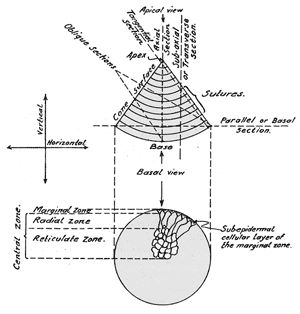
Figure 20: 's "zonation" of discoidal chambers in uniserial-conical shells, namely the marginal, radial and reticulate zones. Compare Fig. 71. From , 1948.
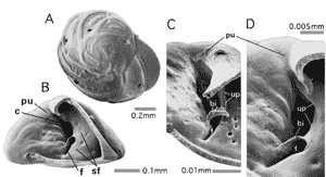
Figure 21: Bipartitor and pseudoumbilicus in Eponides repandus ( et )
from the Gulf of Aqaba, Red Sea, Recent. SEM graphs.
A: dorsal (spiral) view, B: lateral view of dissected specimen. C: oblique umbilical view of dissected specimen. D: detail of rotated oblique umbilical view.
bi: bipartitor; c: spiral umbilical canal; f: foramen; pu: pseudoumbilicus; sf: septal face: note supplementary apertures closed by septal flap covering the septal face; up: umbilical plate.
Figure 22: Biumbilicate shell in Astrononion sp. from the Gulf of Aqaba, Red Sea, Recent. SEM graphs.
A: lateral view; B: apertural view; C: oblique view of dissected specimen showing structural details.
a: aperture; f: foramen; fo: folium; foa: foliar aperture; sf: septal face, covered by septal flap; up: umbilical plate; u: umbilicus, on both sides of the test.
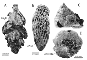
Figure 23: Blades, costae and costellae, linear-elongate ornaments of lamellar-perforate shells.
A: Neouvigerina porrecta (), lateral view;
B: Loxostoma amygdalaeformis (), lateral view;
C-D: Glabratellina sp., lateral and umbilical views. SEM graphs. Gulf of Aqaba. Recent.
Figure 24: Brood chambers in agamonts of
(A-C) Sorites orbiculus () and
(D-E) Amphisorus hemprichii , all from the Gulf of Aqaba. Recent.
A: fresh hatching on empty mother shell under water. B: agamont shell, dried and split open in the equatorial plane. Some embryos remain in situ in the brood chambers. Note size of microspheric proloculus. SEM graph. C: detail of B showing embryo (consisting of proloculus and flexostyle) and surface of resorption including the organic lining. D: equatorial and
E: axial sections showing an abrupt increase in the irregularity and volume of the chamberlet cavity in the last few chamberlet cycles (double arrows) marking the brood chambers. F: Neorotalia sp., thin section parallel to and near the axis of coiling, the last chamber filled with hatchlings. Transparent light micrograph. Spanish Pyrenees, Lower Eocene. G: Orbitolites sp. Oblique-centered thin section, transmitted light micrograph. Note the discrepancy between embryo size and brood chamberlet volumes: we are in presence of either a gamont producing small gametes or zygotes, or of a schizont keeping the offspring in regular broodchambers prior to the first, prolocular shell formation. Lowermost Eocene, Farafrah Oasis, Egypt.
br: brood chamber; e: embryo; ol: organic lining; pr: proloculus; s: septum.
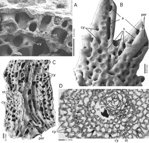
Figure 25: Calyces in arborescent Miniacina miniacea () from the Gulf of Aqaba. Recent.
A: Fragment broken perpendicularly with respect to the axis of the branch, showing an exterior view of the broken funnel walls of the calyces. SEM graph. B: External view of a branch tip showing calyces and apertures at the limiting suture of the expanse chamber that covers previously formed tubular peristomes. SEM graph. C: Fragment broken in the axial plane of branch. SEM graph. D: Thin section perpendicular to the stem axis of the shell. Transmitted light micrograph. Calyces cut obliquely produced characteristic undulating intersections.
a: apertures in marginal position, to be transformed into stolons when overgrown by a subseqent expanse chamber; cy: calyx;
per: tubular peristome either terminal or overgrown by subsequent expanse chambers; pr: proloculus; s: marginal suture of expanse chamber; st: stolon.
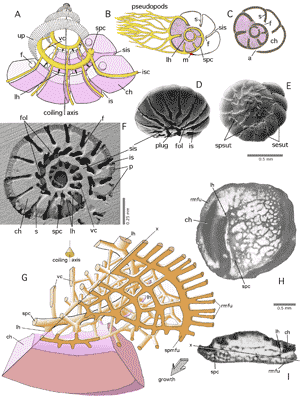
Figure 26: Canal systems.
A: basic geometry of canals in simple rotaliid shells. Schematic, not to scale. Red: chamber plasm; yellow: canal plasm. B-C: during stages of retraction of the chamber plasm into the interior of the shell, the canal system provides motility to the organism by extruding the pseudopods through backdoors, so-called loop-holes. D-F: Challengerella bradyi et alii. Gulf of Aqaba, Red Sea; Recent. SEM graphs. D: oblique-ventral view of umbilical face; E: dorsal (spiral) view; F: Epoxy resin cast of shell cavities showing canal system.
G-H: Hottingerella chouberti (), Northeastern Morocco,
Lower Cretaceous. Schema after , 1976, not to scale, and transmitted light micrographs. Note that the basic geometry with loop-holes in a shell lacking septal subdivision is similar to a rotaliid with chambers.
a: aperture: ch: chamber; f: foramen; fol: folium; is: interlocular, intraseptal space; isc: ingtraseptal canal; lh: loop-hole; m: mask; p: pores; rmfu: radial marginal furrows (open to the ambient environment for their full length prior to being covered by the next whorl); s: septum; sesut: septal sutures; sis: spiral interlocular space; spc: spiral canal; spmfu: spiral marginal furrow; spsut: spiral suture (between successive whorls); up: umbilical plate (general geometric position); vc: vertical canals (at umbilical chamber suture); x: approximate position of section.
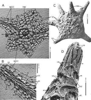
Figure 27: Canaliferous spines in Baculogypsina sphaerulata ( et ), Keij Island, Indonesia. Recent. SEM graphs.
A-B: Epoxy resin casts of shell cavities, the mineralized shell having been dissolved, showing that spine canals originate as extensions of an intraseptal interlocular space, that is then transformed into canals. They feed in their turn the supplemental chamberlets overgrowing the base of the spine. C-D: Shell showing chessboard pattern of supplemental chamberlets, overgrowing the base of the canalled spine and fed in part by the overgrown canal orifices.
a: aperture (of lateral chamberlet); co: canal orifice; csp: canalicular spine; lcl: lateral chamberlet; lh: loop-hole; nep: (spiral) nepiont; sp(c): spine canals; st: stolon.
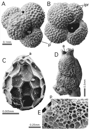
Figure 28: Cancellate ornament in planktic and benthic forms.
A-B: Globoturborotalites tenella (), dorsal view showing supplementary apertures. The cancellate pattern is produced by interpore ridges. C: Favulina hexagona () showing a very regular pattern. D-E: Carpenteria utricularis () showing a more irregular pattern of ridges delimiting areoli with many pores. All from the Gulf of Aqaba. Recent.
a: aperture; ipr: interpore ridge; p: pores; sa: supplementary aperture.
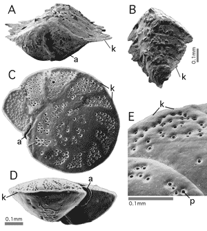
Figure 29: Carina (keel).
A-B: Spiroplectammina cf. taiwanica (), apertural view and side view.
C-E: Paracibicides edomica et , dorsal (spiral) view, peripheral view and detail of perforation pattern along the peripheral carina on the dorsal surface. Note the interiomarginal, extraumbilical - equatorial position of aperture. All specimens from the Gulf of Aqaba. Recent. SEM graphs.
a: aperture; k: carina (keel); p: pore.
The magnification of each figure is in proportion to the length of the 10 cm scale bar above.
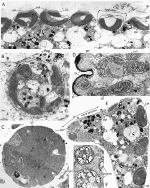
Figure 30: Chloroplasts of foraminiferal symbionts. Gulf of Aqaba. TEM graphs courtesy .
A: Bacillarian (diatom) symbionts below lateral chamber wall, housed in eggholders, below pore mouths, of Amphistegina lobifera . x 5,000. B: dinophycean (dinoflagellate) symbiont of Amphisorus hemprichii . Note sections of short flagellae (arrows) permitting active movement of the symbiont within the lacunar system of the host, to regulate its access to light. The nucleus with its (polyploid) chromosomes visible during the interphase unusual among eucaryotes but characteristic for dinophyceans. x 10,000. C: rhodophycean (red algal) symbiont of Peneroplis planatus ( et ). x 12,000. D: Bacillarian symbiont of Assilina ammonoides (), ultrathin section oblique-tangential to the lateral surface of achamber. Note the loose stacking of the thylacoids in the chloroplast, a characteristic of symbionts of Assilina. x 8,000. E: Elphidium craticulatum ( et ), ultrathin section through a protoplast of a chamber including one of its retral processes (double arrow), showing free chloroplasts in the host chamberplasm, a characteristic of symbiont husbandry. x 4,000. F: Rhodophycean symbionts of Peneroplis planatus ( et ) from Elba in the Mediterranean. Note starch grains covering the pyrenoid and filling most of the symbiont's mass. The starch grains may also appear as free grains in the host chamber plasm and represent seasonal food storage for the host. x 4,000.
bD: basal pore disc; cpl: chloroplast; flag: flagellum with its base; ilD: interlamellar disc of pore; m: mitochondrium; n: nucleus; OL: organic lining; p: pore; Pl: plasmalemma; ppl: pore plug; pyr: pyrenoid; spl: (mineralized) sieve plate; tyl: thylacoid; v: vacuoles.
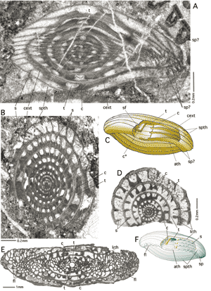
Figure 31: Chomata.
A-C: Eowedekindellina sp. Permian, Iran, collection, oblique, subaxial and subequatorial sections, transmitted light micrographs, and model. Note poleward extension of chomata forming a basal layer. D-F: ordinary chomata in Triticites sp., equatorial and axial sections, transmitted light micrographs and model.
Lower Permian, Spitzbergen. Note in D the sector-wise appearance and disappearance of the chomata due to the slight obliquity of the section.
ath: antetheca; c: choma; c*: cutoff choma stressing the uncertainty about the connection between the choma and the antetheca; fl: fluted septa; kth: keriotheca; s: septum; sp: septal pores; spth: spirotheca; t: tunnel.
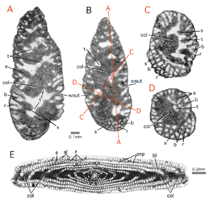
Figure 32: Columellas. Transmitted light micrographs.
A-D: Kurnubia palastiniensis . Upper Jurassic, Northeastern Morocco. A: transverse section near and almost parallel to the axis of coiling; B: axial section; C-D: oblique sections. Position of sections relative to the axial section marked by red lines. E: Borelis schlumbergeri (), axial section. Gulf of Aqaba. Recent. Note double columella in planispiral-fusiform shells.
b: beam; bl: basal layer; col: columella; f: foramina; prp: preseptal passage; r: rafter; s: septum; sl: septulum; t: tunnel; wsut: whorl suture.
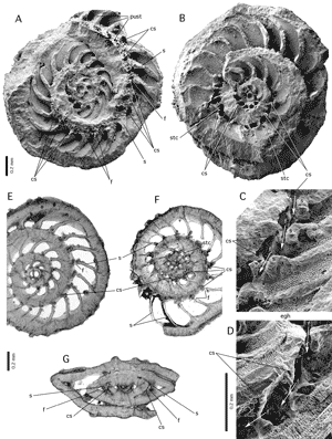
Figure 33: Countersepta.
A-D: Amphistegina tuberculata , Dominican Republic,
Upper Miocene, SEM stereographs showing the split halves of a megalospheric shell. A: dorsal side in ventral view, B: ventral side in dorsal view, with details in C and D.
E-G: Amphistegina lopeztrigoi , thin slides in transmitted light micrographs, Eocene, Florida. Generic and specific name are unclear and need revision. E: centered section perpendicular to the coiling axis; F: section perpendicular to the coiling axis located below the megalosphere; G: poorly centered axial section.
cs: counterseptum; egh: eggholder; f: main foramen; pust: pustules covering successive faces; s: septum of main chambers; stc: stellar chamberlets.
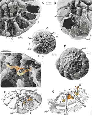
Figure 34: Cover plate and foramenal plate in Challengerella bradyi et alii from the Gulf of Aqaba. Recent. SEM graphs.
A-B: dissected specimens in oblique ventral view. C: young specimen, ventral view. Note incipient umbilical pile against which the foliar walls are beginning to fuse, covering the spiral interlocular space. D: dorsal (spiral) view of adult megalospheric specimen. E: Detail showing foraminal and cover-plates. Colours corresponding to those used in the models. White arrow: loop-hole. F: general model showing position of plates and communications. Note position of loop-hole (red arrow). G: Detail of umbilical structures in last three chambers. Note the separation of streaming chamber plasm through successive foramina (blue arrow) and circulation in the umbilical interlocular space (black arrow) by the umbilical structures. Models schematic, not to scale, after , 2000.
a: aperture; cp: coverplate; csut: chamber (septal) suture; f: foramen; fo: folium; foa: foliar aperture; fp: foramenal plate; isp: interlocular (intrasepal) space; lh: loop-hole; n: notch; pil: umbilical plug; s: septum; sf: septal flap; suc: spiral umbilical interlocular space, transformed into spiral canal when covered by foliar extensions; u: umbilicus; ulch: ultimate chamber; vc: funnel (vertical canal); wsut: whorl (spiral) suture.
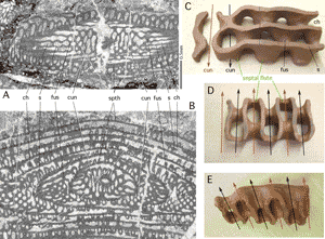
Figure 35: Cuniculi and septal fluting in Eopolydiexodina sp. Transmitted light micrographs. collection, Permian, Iran.
A: shallow tangential section parallel to coiling axis. B: Deep transverse section parallel to coiling axis. C-E: plasticine model sculptured about 1945 by (* 1896 - † 1984). C: Oblique peripheral view showing undivided peripheral parts of two successive chambers. Arrows point in the direction of growth. B: Proximal view showing cuniculi (arrows). C: oblique proximal view showing fluted septal face and cuniculi.
ch: chamber lumen; cun: cuniculus; fus: point of fusion of subsequent septal flutes in opposing positions; s: septum; spth: spirotheca carrying a keriotheca.
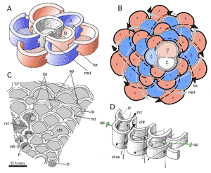
Figure 36: Development of chamberlet cycles from an auxiliary chamber in an early orbitoid, the Campanian Lepidorbitoides minima from Mexico, after et alii, 2002.
A: stereograph of a megalospheric embryo consisting of a biconch (protoconch and deuteroconch) supplemented by a third chamber (auxiliary chamber) with a median and two lateral stolons. Successive growth stages coloured alternately in red and blue. Lamellation omitted. Schematic, not to scale. B: development of chamberlet cycles. Schema, not to scale. Inner lamella omitted, outer lamella alternating in white or plain black in successive growth stages. Axes of successive stolons form overcrossing spirals (arrows). Median stolons indicated by dotted arrows, lateral stolons by black arrows. C: Camera lucida drawing of a slightly oblique section of the equatorial main chamberlet layer of adult growth stage showing retrovert apertures and annular passages. D: stereograph of adult chamberlets of two successive cycles with their crosswise-oblique stolon axes, the annular passage and their retrovert stolons. Schematic, not to scale.
A: auxiliary chamber; D: deuteroconch; P: protoconch; X: closing chamberlet filling up the first chamberlet annulus; ap: annular passage; chl: chamberlet lumen; lst: lateral stolon; mst: median stolon; p: pores; rst: retrovert stolons; stax: crosswise-oblique stolon axes.
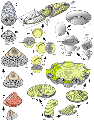
Figure 37: Chamber arrangement and apertural face. Schematic, not to scale. In part after , 2000.
1: planispiral-spiroliniform. 2: planispiral-evolute and flaring, peneropliform. 3: planispiral-evolute, approaching reniform, 4: annular-concentric, with thickened and folded margins, as in Marginopora vertebralis. 5: planispiral-involute, as in Archaias. 6: biserial � textulariid. 7: biserial-cuneiform. 8: uniserial-conical. 9: uniserial-conical with marginal apertures. 10: streptospiral-involute as in Pseudonummuloculina. Coiling axis rotating with each chamber. 11: streptospiral with planispiral-involute adult stage, as in Helenalveolina. 12: planispiral involute. Coiling axis fixed throughout ontogeny. 13: planispiral-fusiform: A: axial section, E: equatorial section. Black arrow: direction of growth; white arrow: direction of movement. 14: miliolid (-quinquelocular, -trilocular), with fixed apertural axis and with coiling axis rotating in perpendicular position in respect to apertural axis. 15: miliolid-bilocular with fixed apertural and coiling axes. 16: unilocular-concentric, with discoidal trematophore, as in Lacazina elongata. 17: unilocular-concentric with annular trematophore, as in Lacazina compressa. 18: low-trochospiral, as in Rotorbinella. 19: high-trochospiral, as in Sakesaria.
a: aperture; af: apertural face; apax: apertural axis; col: columella; fol: folium; ma: marginal aperture; n: notch; per: periphery; potort: polar totion; pst: peristome; spax: coiling axis.
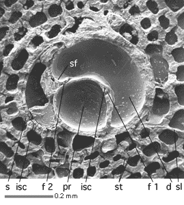
Figure 38: Deuteroconch in Cycloclypeus carpenteri (), Bikini, Pacific. Recent. SEM graph of shell split open in the equatorial plane.
d: deuteroconch; f 1: foramen of protoconch; f 2: foramen of deuteroconch; isc: intraseptal canal system; pr: proloculus; s: septum; sl: septulum; st: stolon (Y-shaped).
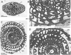
Figure 39: Diaphanotheca in Fusulina distenta et , Hartville
Fm., DesMoines series, Guernsay, Wyoming, Upper Carboniferous. Transmitted light micrographs.
A: slightly oblique, imperfectly centered axial section; B: detail of A showing diaphanotheca in axial section of adult whorls; C: equatorial section showing diaphanotheca extending over septal faces; D: detail of other specimen.
c*: choma; dth: diaphanotheca; pr: megalosphere. Note presence of some kind of flexostyle. s: septum; t: tunnel.
The magnification of each figure is in proportion to the length of the 10 cm scale bar above.
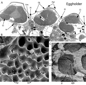
Figure 40: Eggholders harbouring the symbionts in Heterostegina depressa d'. Gulf of Aqaba. Recent.
A: TEM micrograph of section perpendicular to lateral chamber wall; biomineralized wall dissolved, organic cell walls distinctly separated by the techniques of preparation. Courtesy . x 10,200. B-C: dried shell, split open in the equatorial plane. The internal surface of the lateral chamber wall bears eggholders. SEM graphs, x 5,000 and x 500. Drying strechted the organic cell walls.
Cl: chloroplast of bacillariophycean symbiont; clat: lateral intraseptal canal; egh: eggholder; M: mitochondria; OL: organic lining of host; Pl: plasmalemma of host; Plp: pore plug (see pore); Pls: plasmalemma of symbiont; Py: pyrenoid; s: septum; sl: septulum; st: stolon (Y-shaped); V: Vacuole housing symbiont; Vm: vacuolar membrane.
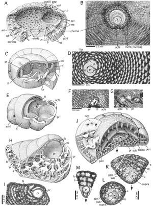
Figure 41: Embryonic apparatus.
A: Discocyclina, stereograph after (1999), modified. Schema, not to scale. B: Discocyclina sp., equatorial section, showing corona. Transmitted light micrograph. C: Amphisorus sp. Stereograph, schema, not to scale. D: Amphisorus hemprichii from the Gulf of Aqaba, Red Sea. Recent. Equatorial section, transmitted light micrograph, showing flexostyle and forecourt. E: Orbitolites sp., stereograph after , 1961, redrawn. Note the shape like a dumb-bell of the proloculus. F-G: Orbitolites sp., Lower Eocene, Pakistan, collection. Oblique-centered sections, in approximately equatorial and axial direction, showing flexostyle constricting the proloculus. Transmitted light micrographs. H: Orbitopsella sp., stereograph showing sphaeroconch with its exoskeleton. Schema, not to scale, after , 1967. Note the appearance of endoskeletal pillars not before the third chamber. I: Orbitopsella praecursor (), Middle Lias, Morocco, equatorial section showing the thin proloculus wall and the sphaeroconch with its simple exoskeleton consisting of beams only. Transmitted light micrograph. J: Advanced orbitoliniform embryo, schematic, not to scale. Note the apex that is directed downwards (arrows) to facilitate the comparison with 's drawings (1963). K-L: embryos of Karsella hottingeri , a
Upper Paleocene form that exhibits an architecture strikingly similar to the one of Mid-Cretaceous orbitolinids. Pakistan, collection. Transmitted light micrographs. M: Sabaudia minuta Jr, embryo followed by three agglutinated chambers of the same series and presenting a simple exoskeleton.
Lower Cretaceous. Note the hyaline embryo wall. The embryos are known to be formed within the mother shell and seem to have no access to grains in the environment to build their agglutinated walls.
achl: auxilliary chamberlet; ap: annular passage; ast: annular stolon; b: beam; d: deuteroconch; ecr: equatorial crest; fl: flexostyle; il: inner lamella; lchl: lateral chamberlet; mchl: main chamberlets; ol: outer lamella: ost: oblique stolon; ost(d): oblique stolon of deuteroconch; p: pore; pap: papilla; peri: periembryonal chamberlets; pi: pillar; pr: proloculus; rst: radial stolon; rst(d): radial stolon of deuteroconch; s: septum; sc: sphaeroconch; sl: septulum; sub: subembryonic chamber (= deuteroconch); supra: supraembryonic chamber.
Figure 42: Epiphytic foraminifera and trichomes. Recent. Gulf of Aqaba, Red Sea. SEM graphs.
A: Epiphytes on Halophila leaf. Pl: Planogypsina acervalis (); Ha: surface of Halophila leaf with leaf hairs (trichomes); So:
Sorites orbiculus (). B: Trichome on Halophila leaf and aperture in a radially-marginal position of the epiphytic Planogypsina acervalis (). C: Opened test of Planogypsina acervalis showing the inner side of the perforate, dorsal chamberlet walls, coating the overgrown trichomes of their substrate, and the dorsal part of the septal walls with stolons representing intercameral foramina.
a(m): aperture of a marginal, terminal chamberlet; chl(m): chamberlet of the ultimate marginal chamberlet cycle; dw(chl): dorsal wall of chamberlet; ec: epithelial cells of the Halophila substrate; p: pore; st: stolon; tri: trichome.
Figure 43: Endosolen in Favulina melosquamosa (), Gulf of Aqaba. Recent. SEM graphs.
A: dissected specimen, oblique aboral view; B: lateral view; C: aboral view.
a: aperture; et: endosolenian tube.
Figure 44: Enveloping canals produced by folded outer lamellas.
A-D: Lamellae in the peripheral portion of three successive chambers. Stereograph, schematic, not to scale. A: ultimate and penultimate chamber: red: inner lamella; green: outer lamella of ultimate chamber, white: outer lamella of penultimate chamber. B: addition of the next chamber with an inner lamella in red and an outer lamella in blue. C: addition of an other chamber with its outer lamella in yellow. D: superposition of lamellas B and C over the then final chamber A after two additional growth steps. E: SEM micrograph of the complete test of Calcarina defrancii d', ventral view, showing distribution of canal orifices over all the test including the canaliferous spines. F: SEM micrograph of an epoxy resin cast of the shell cavities in Calcarina gaudichaudii d' cut in a direction perpendicular to the axis of the spiral shell, ventral view; G: detailed SEM micrograph of the ventral shell surface of C. gaudichaudii. Calcarinas from Keij Island, Indonesia. Recent. After and , 1980.
apil: axial pile of lamellae; ch: chamber and chamber lumen; co: canal orifice; csp: canaliferous spine; envc: enveloping canal; isc: intraseptal space or canal; il: inner lamella; lh: loop-hole; ol: outer lamella; p: pore; paf: perforate part of apertural face; ssk: supplemental skeleton; st: stolon; uc: umbilical canal network; uch: ultimate chamber suture, chamber walls broken off.
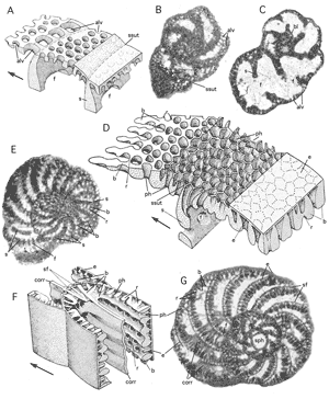
Figure 45: Alveolar exoskeleton and polygonal network.
A-C: simple alveolar layer in Everticyclammina virguliana (), Mechra Klila, Northeastern Morocco, Uppermost Jurassic. A: stereograph, schematic, not to scale. B: tangential section. Note the large size of the alveoles in a postseptal position. C: para-equatorial, non-centered section showing septa and the basal coat at the bottom of the chamber, resembling a basal layer. E-G: polygonal network in Spirocyclinidae. E: Choffatella tingitana , megalospheric generation, in tangential section near to the equatorial plane. Note the clear differentiation of beams and rafters. D: stereograph of spirocyclinid polygonal network. Note the curved pigeon holes in preseptal position which in axial section might be mistaken for foramina. F-G: extension of beams into a corrugated sheet that replaces endoskeletal pillars in Hottingertidae. F: stereograph representing a part of an axial section. Not to scale. G: Alveosepta powersi (), Northeastern Morocco, Upper Jurassic. Equatorial section of megalospheric specimen.
alv: alveoles; b: beam; bl: basal layer; corr: corrugated median extension of beams; f: foramen; ph: pigeon holes; s: septum; sf: supplementary foramina; sph: sphaeroconch; ssut: septal suture. Arrows: direction of growth. After , 1967.
Figure 46: Expanse chambers and calyces.
A: Sketch of ultimate and penultimate expanse chambers, not to scale. B: A
cut-out portion of five successive expanse chambers, cut in an oblique direction
with respect to the shell axis, schematic, not to scale. Lamellation of walls omitted. C: Ultimate and penultimate expanse chambers in Planogypsina squamiformis (). Gulf of Aqaba, Red Sea; Recent.
SEM graph. D-G: Expanse chambers with calyces in Miniacina miniacea (), Gulf of Aqaba, Red Sea; Recent.
D-F: SEM graphs, G: thin section in transmitted light. D: broken surface perpendicular to the axis of a branch of the arborescent
shell: the expanse chambers are grouped around tubular extensions of the peristomes of earlier chambers. E: a fragment of shell showing
the inner surfaces of two successive expanse chambers. Note the alternating arrangement of the calyces. F: radial spreading of ultimate expanse chamber at the foot of an arborescent shell,
thus enlarging the shell's surface of attachment. G: centered section near and parallel to the surface of attachment, so cutting the latest, outermost expanse chambers at diminishing angles.
a: aperture; ax: shell axis; cy: calyx; p: pore; pr: proloculus; puch: penultimate expanse chamber; sut: suture of expanse chamber; tpa: tubular passage connecting parts of expanse chamber; uch: ultimate expanse chamber.
Figure 47: Endoskeletal patterns in discoidal shells.
A-D: disposition of apertural axes. A: radial axes alternating in radial position from one stolon layer to the next. This is the most common disposition in imperforate forms with annular stages of growth. B: radial axes superposed in radial position on all stolon planes. C: crosswise-oblique stolon axes alternating in radial position from one stolon plane to the next. D: crosswise-oblique stolon axes superposed on all stolon planes. This pattern characterizes all members of the orbitolitid family. Schematic, not to scale. E-H: all endoskeletal elements
are disposed in accordance with the basic patterns of the foraminal axes: E corresponds to pattern A, F to pattern B, G to pattern C and H to pattern D. Stereographs after , 1967. Schematic, not to scale. In reality, the patterns are often disturbed by intercalary
elements generated as the diameter of the annuli increases during growth. This
maintains on the apertural face the mean distances between apertures and their mean diameter constant during ontogeny. Examples: E1-2: Pseudotaberina malabarica, megalospheric generation, from Iran. Middle Miocene. E1: oblique-centered section of spiral-involute stage showing radial disposition of pillars. Laterally, there is a layer of short septula. E2: a transverse section tangential to a septum shows the alternating disposition of the foramina and the pillars. F1-2: New genus (possibly related to Pastrikella) from the Pyrenean
Upper Cretaceous in Northern Spain. The endoskeleton consists
only of septula. There is but one median annular preseptal passage and it occupies the total radial extension of the annular chamber.
There are only two planes of stolons. F1: an oblique section at a low angles
with respect to the equatorial plane shows the radial disposition of the apertural axes and of the septula. F2:
a transverse section parallel to the shell axis shows that the stolon axes on the two stolon
planes are superposed. G1-3: Amphisorus from Rottnest Island near Perth, Australia. Recent. G1: the detail of an equatorial section demonstrates the crosswise-oblique disposition of the pillars on neighboring stolon planes.
G2, an equatorial section, demonstrates that pillars are restricted to the equatorial zone of the disc. G3:
a transverse section parallel to the shell axis and tangential to an annular septum shows the disposition of the median foramina and pillars altermating in radial position on successive stolon planes. They are flanked by two annular preseptal passages separating them from a lateral layer of septula subdividing the annular chamber. H1-2: Orbitolites spp. from the region of Tremp, Lerida prov., Northern Spain. Pyrenean Lower Eocene (Ilerdian). H1: the comparatively regular disposition of the ramps in sections parallel to the equatorial plane
reveals their superposition in consecutive stolon planes. H2: in the transverse section parallel to the axis of the shell this superposition is clearly visible where the section is tangential to an annular septum.
Abbreviations: b: beam; f: foramen; pi: pillar; prp: preseptal space; ra: ramp; s: septum; sl: septulum.
Figure 48: Faces (coloured red) in biserial and spiral forms. SEM graphs (if not specified otherwise) of specimens from the Gulf of Aqaba, Red Sea. Recent.
A-C: Textularia aff. goesi , apertural and lateral views of intact specimens and frontal view of broken specimen showing septal face. Note the smooth surfaces characteristic
of the faces of many agglutinated forms. D-E: Discorbinoides sp. A in et alii, 1993. Plastogamic pair and plastogamic umbilical face with radial grooves. F-G: Glabratellina sp. A in et alii, 1993. Ventral view showing plastogamic ventral face with radial grooves and lateral view showing shell whorls with their sutures. H-I: Bolivinella elegans , a lamellar-perforate, biserial form with known plastogamic reproduction,
that has radial grooves on its face. J: Floresina spicata ( et )
has a smooth face with few radial grooves and in addition grooved septal sutures. A
(plastogamic?) plate covers the narrow umbilicus. As yet, plastogamy has not been observed in vivo in this species. K-L: Amphistegina lobifera , Megalospheric specimen in
incident light micrograph, ventral view and detail of apertural face. Note the extension of the ornamented surface
for a notable distance over peripheral-ventral parts of the previous whorl. M-N: Amphistegina bicirculata , megalospheric
specimen in incident light micrograph, ventral view and detail of apertural
face. Note comparatively much smaller ornamented surface corresponding to lower water energy in its deeper habitat.
a: aperture; af: apertural face; f: foramen; p: pore; plp: plastogamous (?) plate; pp: parapore; rgr: (plastogamous) radial grooves; sfa: septal face; stch: stellar chamberlet; stsut: stellar suture; sut: (chamber) suture; wsut: whorl suture.
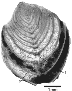
Figure 49: Equitant chambers in Frondicularia sp. from the Prerif, Northwestern Morocco, Lower Miocene. Transmitted light micrograph of section in the sagittal plane of a microspheric specimen. Note the denticulate margin of the single, stellar foramen in terminal position (f) and the extremely fine perforation of the septal wall that is characteristic of most nodosariids.
Figure 50: Feathered
grooves. All specimens figured as SEM graphs.
A-B: Ammonia beccarii (), Adriatic Sea, Recent. Peripheral and oblique - umbilical views. Note the ridges on the apertural face that initiate the feathering when the next chamber is added during ontogeny. C-D: Neorotalia calcar (d'), Keij Island, Indonesia. Recent. Ventral view of an entire specimen and detail of a ventral feathered groove covered by a folded outer lamella. Note the imperforate nature of the cover and the orifices of the canal system. Compare: enveloping canal systems in Fig. 44. E-G: Unfeathered grooves in Ammonia ikebei ( et ), Eastern Kalimantan, Borneo,
Upper Miocene. Peripheral view of entire specimen and details in oblique-umbilical and in dorsal views. Note the hemicircular attachment of the septal flap and the undivided umbilical plug that correspond to unfeathered septal
grooves.
af: apertural face; f: foramen; fea: feathered grooves; ol: (folded) outer lamella covering a septal groove that is converted to an interlocular space; or: canal orifice; pil: pile; s: septum; sgr; septal groove (unfeathered); spgr: spiral groove (unfeathered); upl: umbilical plug.
Figure 51: Fistulose chamberlets. All specimens from the Gulf of Aqaba, Red Sea, Recent.
A-C: Sahulia kerimbaensis (). A: Detail of an edge view, SEM graph; B: side view, X-ray graph, black background
removed. Note the narrowness and the irregularity of the fistulose chamberlets in this species. C: edge view, SEM graph. D-H: Spirotextularia floridana (). D: paraporous partition separating the chamber lumen from a fistulose chamberlet. SEM graph. E: Detail of sectioned paraporous partition. SEM graph. Note the organic lining that closes off the parapores from the chamber lumen. SEM graph. F: Shell fragment broken in a direction perpendicular to the shell axis, showing paraporous partition between the chamber lumen and the fistulose chamberlet. SEM graph. G: Lateral view, X-ray graph. H: lateral view of an entire specimen. SEM graph. Note the spiral arrangement of the nepionic chambers.
a: aperture; af: apertural face; ch: chamber lumen; f: foramen; fis: fistulose chamberlet; OL: organic lining; pp: parapore; ppa: paraporous partition; pr: proloculus; pv: pavement; s: septum.
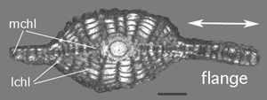
Figure 52: The flange (double arrow) in Nephrolepidina sp., axial section, transmitted light. Oligocene, Sarawak, Borneo.
lchl: lateral chamberlets; mchl: median chamberlet layer. Scale bar 0.2 mm.
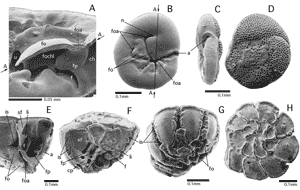
Figure 53: The folium and its apertures. All specimens from the Gulf of Aqaba, Red Sea, Recent. SEM graphs.
A-D: Rosalina bradyi (). A: detail of dissected specimen, oblique-ventral view. The approximate position of the breakage surface (arrows A-A) is indicated by the line A-A in Fig. B. B: ventral view showing the folium
at its maximum development, with anterior and posterior apertures. C-D:
peripheral and dorsal views. Note the restriction of the perforation to the dorsal surface of the shell, an indication that the face extends from the umbilical side of the shell over its periphery. E-H: Asterorotalia gaimardi (d'). E-F: dissected specimens showing details of advanced umbilical architecture covered by the folia: foraminal and coverplates (compare Fig. 34). G: oblique-ventral view showing the folia that cover the ventral part of the interlocular space. H: dorsal view showing the spiral arrangement of the chambers.
a: aperture; ch: chamber lumen; cp: coverplate; f: foramen; fo: folium; foa: foliar aperture; fochl: foliar chamberlet lumen; fp: foraminal plate; is: interlocular space; li: lip (of foramen); n: notch; sf: septal flap; u: umbilicus.
Figure 54: Fossette, parafossette, ponticulus and retral process in advanced elphidiids from the Gulf of Aqaba, Red Sea. Recent. SEM graphs except D and H.
A-F: Elphidium striatopunctatum ( et ). A: lateral (umbilical) view of the surface of the shell. B: detail showing the orifices of the canal system. Note the heavy spiking of the orifices, a device to fend off
inedible particles such as diatom frustules. C: the detail of an orifice of a parafossette. Note its alignment with the axis of the adjacent ponticulus. D: axial section of megalosperic specimen, transmitted light micrograph. Note the row of foramina at the base of the chamber and the row of retral processes in the
chamber roof. E: epoxy resin cast of the cavities of the shell showing the narrow spiral of the umbilical canal and the double row of fossettes and parafossettes representing the septal interlocular space. F: detail of a cast showing the disposition of fossettes and parafossettes and their connection to the intraseptal canal system.
G-L: Elphidium craticulatum ( et ). G: oblique-peripheral view of entire shell. H: axial section of megalospheric specimen. Transmitted light micrograph. Note the single row of foramina at the bottom of the chamber and the broad umbilical pile with its funnels. I: apertural face showing the masked apertures. Note the imperforate keel, an important specific character. J: detail of G: the orifices of the fossettes in the last chamber alternate with tiny parafossettes. These disappear when they are covered by subsequent secondary lamellation in later stages of growth. K: epoxy resin cast showing umbilical cavities including the foliar chamberlets at the tips of the alar prolongation of the chamber. The foliar chamberlets are transformed during ontogeny into a spiral umbilical canal. L: epoxy resin cast showing detail of septal architecure: the alternation of retral processes and fossettes. Note the tiny pores on the cast of the main chamber lumen and the traces of the spikes on the walls of a fossette.
a: aperture; af: apertural face; f: foramen; fo: folium; foa: foliar aperture; foc: foliar chamberlet; fs: fossette; k: keel; m: mask; mc: main chamber lumen; pfs: parafossette; po: ponticulus; rp: retral process; s: septum; suc: spiral umbilical canal; upil: umbilical pile; vc: vertical canal (funnel). Scale bars: 0.1 mm, double scale bars: 0.05 mm.
Figure 55: An umbilical architecture dominated by funnels: Dictyokathina simplex , megalospheric specimens from
Qatar, Paleocene. Transmitted light micrographs.
A: subaxial section showing funnels in their longitudinal extension. B: transverse section near the axis of the shell with the positions of sections A &
C-D indicated. C-E: sections more or less perpendicular to the axis of the shell showing the vast umbilical area crowded with funnels. Note the chamber arrangement in multiple spirals, The start of supplementary spirals is indicated by arrows. The material does not allow
resolution of the question: is the first chamber of a supplementary spiral fed from a foramen or from a
canal?
ch: chamber lumen; f: foramen; fu: funnel (vertical canal); isp: intraseptal interlocular space; ssp: spiral interlocular space; up: umbilical plate.
The magnification of the figure is in proportion to the length of the 10 cm scale bar above.
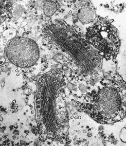
Figure 56: The Golgi apparatus and mitochondria in the cytoplasm of Alveolinella quoyi (d') from the
Maldive Islands, Indian Ocean. Transmission electron micrograph, x 50,000. Courtesy .
G: Golgi apparatus with its cisternae (cist); M: mitochondrium with its cristae (cr).
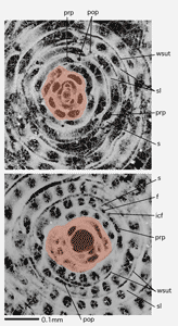
Figure 57: Glomerulus (coloured) in Glomalveolina lepidula () from Tremp, Northern Spain, Ilerdian. Approximately equatorial section with small, ?microspheric proloculus and axial section with large, ?megalospheric proloculus. Incident light.
f: foramen; icf: intercalary foramen; pop: postseptal passage; prp: praeseptal passage; s: septum; sl: septulum; wsut: whorl suture (enlarged by slight deformation).
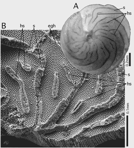
Figure 58: Hemiseptula in Amphistegina lessonii d' from the
Gulf of Aqaba, Red Sea. Recent.
A: the dorsal surface of a megalospheric specimen. Incident light. B: the inner surface of a fragment of dorsal chamber walls showing septa and hemiseptula broken at
a short distance from the level of the eggholders. Note the three-layered texture of the septa (a primary
bilamellar wall plus a septal flap) and the two-layered nature of the hemiseptula that results from the folding of the inner lamella. SEM graph.
egh: eggholder; hs: hemiseptula; s: septa.
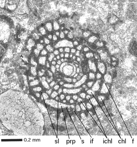
Figure 59: Intercalary chamberlets in Borelis curdica (). Qum
Basin, Iran. collection 229. Lower Miocene. Axial section.
chl: chamberlet; f: foramen; ichl: intercalary chamberlet; if: intercalary foramen; prp: preseptal passage; s: septum; sl:
Y-shaped septulum.
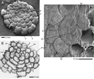
Figure 60: Surface of attachment with supplementary apertures in Planorbulina mediterranensis d'. Elba Island, Mediterranean. Recent.
A: Free surface with apertures in subperipheral position. SEM graph. B: Equatorial section showing the nepionic, keeled spiral chambers followed by early chamberlet cycles with their oblique foramina and supplementary apertures where the section passes immediately below or
within the attached chamber walls. Note the straight septa between the proconch
and the two following chambers forming together a triconch. Transmitted light micrograph. C: Detail of surface of attachment with its tiny supplementary apertures in sutural position. Note the keeled nepiont. SEM graph.
a: aperture; f: foramen; k: keel of the nepiont; p: pore; pr: proloculus; sa: supplementary aperture.
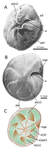
Figure 61: Infundibulum and marginal prolongation.
A: Neoeponides bradyi (), oblique-ventral view showing infundibulum.
SEM graph; Gulf of Aqaba, Red Sea; Recent.
B: Eponides repandus ( et ), ventral view showing marginal prolongation.
SEM graph; Gulf of Aqaba, Red Sea; Recent.
C: Schematic drawing showing position of marginal prolongations in respect to ventral and dorsal test morphology in the last whorl of Eponides repandus.
Gray: dorsal view with raised sutures; red: outline of chamber walls at level ventrally below periphery; green: ventral ouline of chambers below level of marginal prolongation.
a: aperture; inf: infundibulum; mpr: marginal prolongation; per: (imperfotate) periphery; s(d): (oblique) septum (as seen in dorsal view); s(per): position of septum at level of periphery; ss(v): (radial) septal suture in ventral view.
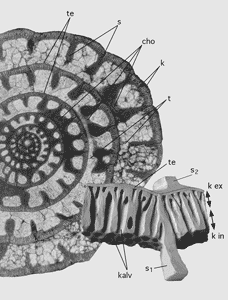
Figure 62: Keriotheca (double arrow) in equtorial section of unidentified Triticites and as plasticine model by (unpublished, modified).
cho: choma; kalv: keriothecal alveoles; k ex: extern keriotheca; k in: intern keriotheka; s: septum; s1: septum belonging to whorl bearing the keriotheka; s2: septum of following whorl; te: tectum.
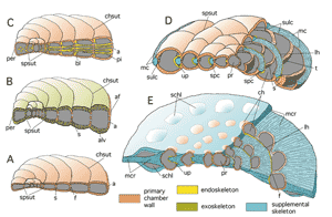
Figure 63: Comparison of foraminiferal skeletons. Schematic, not to scale. Lamellation, perforation and canal orifices omitted.
A: a planispiral-evolute shell without skeletal structures, composed of simple primary chamber walls with multiple apertures, such as
that of Peneroplis. B: a planispiral-evolute shell with an alveolar exoskeleton, such as Pseudocyclammina. C: a planispiral shell
with a pillared endoskeleton such as Archaias. Note that in the axial sections of shells with peneropliform, flaring chambers the periphery of the shell and the
apertural face are on opposite sides. Consequently, the pillars extending from chamber bottom to chamber roof appear in the axial plane
on the side cutting the apertural face as longitudinal and on the other side
cutting the periphery as more or less perpendicular sections. D: a spiral shell with a supplemental skeleton restricted to the periphery of the shell, as in nummulitids
with a marginal cord. E: a spiral shell with an enveloping canal system and a marginal crest as in Pellatispira. Note the primary lateral chamber walls "emerging" from the supplemental skeleton. These primary chamber walls are covered by secondary lamellae but
are perforated in continuation of the primary bilamellar wall. Therefore they
are not a part of the supplemental skeleton. The supplemental chamberlets have perforate lateral walls but do not communicate directly with the spiral chambers by retral stolons. They are fed by canal orifices.
a: aperture; af: apertural face; alv: alveole; bl: basal layer; ch: chamber; chsut: chamber suture; f: foramen; lh: loophole; mc: marginal cord; mcr: marginal crest; per: periphery; pi: pillar; pr: proloculus; s: septum; schl: supplemental chamberlet; spc: spiral canal; spsut: spiral suture; sulc: sulcus; t: tunnel; up: umbilical plate.
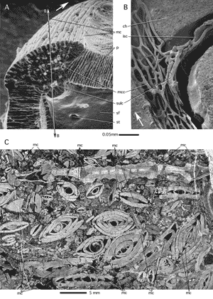
Figure 64: Marginal cord in nummulitids.
A-B: Assilina ammonoides (), Recent, Gulf of Aqaba, Red Sea. SEM graphs after , 1977. A: Detail of marginal cord in
a shell broken in the axial plane. Double arrow B - B indicates the approximate position of
the cut in the cast of Fig. B. B: Detail of marginal cord in an epoxy resin cast
that was cut approximately in the equatorial plane (as indicated in Fig. A), showing canal and chamber lumina after dissolution of shell. White arrows indicate direction of growth. C: Nummulitic limestone from Steinbach, Einsiedeln, Swiss Helvetic Alps, Lower
Eocene (Cuisian). Transparent light micrograph of section perpendicular to bedding plane. 1: Assilina sp. of A.
ammonea- praespira phyletic line, with particularly large and prominent marginal cord; 2: Nummulites spp.; 3: Assilina sp. of A. spira phyletic line; 4: Assilina sp. of A. exponens phyletic line; 5: Asterigerina cf. rotula ():
no marginal cord.
ch: chamber lumen; isc: intraseptal canal system; mc: marginal cord; mcc: marginal cord canals; sf: septal flap; st: stolon; sulc: sulcus canal.
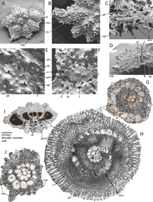
Figure 65: Supplemental skeletons including marginal crests.
A-G: Pellatispira group provalei . H: P. fulgeria . Both species from Kalimantan, Borneo, Indonesia.
Middle-Upper Eocene. I-J: Calcarina sp., Kutei basin, Kalimantan. Pleistocene. A-D: SEM micrographs. G-J: transmitted light micrographs of oriented thin sections of free specimens.
A: the free nepiont shows early spiral chambers not yet covered by a supplemental skeleton. An
uncovered open interlocular space remains between the ultimate and penultimate chambers. B: detail of lateral view of a free nepiont revealing the early presence of canaliculate spines in the first volution of the spiral chambers and the modest
extent of the marginal crest at this stage of growth. C: peripheral view of the margin of the second whorl. Note the strong radial spikes that support the thin imperforate walls of the marginal crest. D: septal face in oblique-peripheral view: the septal flap is reduced to a small area above the foramen. E: a lateral, flying cover of the interlocular space, with canal orifices at its margins, is produced by a free fold of an outer lamella and represents thus a primary element of the supplemental skeleton. F: in later growth stages, a first imperforate cover of the interlocular space may be bridged by supplementary chamberlets with a perforate, bilamellar wall. G: equatorial section. The primary bilamellar walls of the spiral chambers are coloured. All uncoloured
constituents of the shell are part of the supplemental skeleton. H: Extreme development of the supplemental skeleton as
a broad marginal crest covered with piles that are flanked by the canals of an enveloping system. I: the axial section of a trochospiral shell demonstrates the complex pattern of
the umbilical cavities between umbilical piles of lamellae. The primary bilamellar walls of the spiral chambers are
coloured. J: a section perpendicular to this axis of coiling shows that canalicular spines grow outward from the supplemental skeleton that envelops the primary bilamellar (coloured) wall of the spiral chambers.
Abbreviations: a: aperture; c: canals, canal orifices; ch: (spiral) chamber; chl: (supplemental) chamberlet; csp: canaliculate (pseudo)spine; f: foramen; is: intraseptal interlocular space; lh: loophole; mcr: marginal crest; p: pore; pil: pile (of lamellae); pr: proloculus; s: septum; schl: supplemental chamberlet; sf: septal flap; sk: supplemental skeleton; spi: spike; uc: umbilical cavity system; up: umbilical plate.
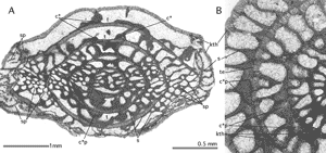
Figure 66: Septal and chomatal "pores" in Triticites plummeri, Graham Fm., Texas, Permian. Transmitted light micrographs.
A: subaxial section; B: details in equatorial section. Both pore-like features have nothing in common with true pores. Septal pores are tiny multiple
foramina. The nature of chomatal pores, a wall texture particular to fusulinids, is not understood at present.
c: choma; c*p: chomatal "pores"; kth: keriotheka; sp: septal "pores". t: tunnel; te: tectum.
The magnification of the figure is in proportion to the length of the 10 cm scale bar above.
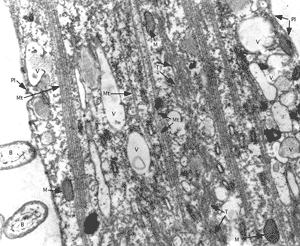
Figure 67: Microtubules (Mt) and tubulin paracrystals (T) in canal ectoplasm of Assilina ammonoides (). TEM micrograph, courtesy , x 24,000.
B: bacteria; M: mitochondria; Mt: microtubule; Pl: plasmalemma; T: tubulin paracrystals; V: vacuoles with or without fibrillar content (waste products?).
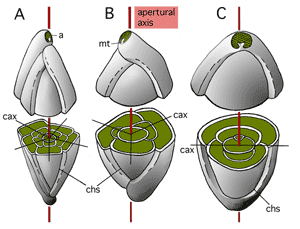
Figure 68: Milioline coiling.
Schematic, not to scale. Note the coiling mode: only two chambers per whorl. Chamber lumina green, coiling axes black.
Foramina aligned in the apertural axis (red).
A: quinqueloculine mode of coiling: for each new chamber the coiling axis rotates 72° around the apertural axis. B: triloculine mode of coiling:
for each new chamber the coiling axis rotates 120°. C: biloculine mode of coiling: the coiling axis does not rotate.
a: aperture; cax: coiling axis; chs: chamber suture; mt: miliolid tooth.
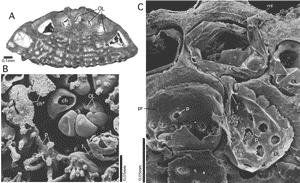
Figure 69: Organic lining.
A: Lockhartia haimei (), megalospheric specimen, axial section, transmitted light micrograph. From the Salt Range, Pakistan. Paleocene. Note the detachment of the organic lining from the inner surface of the biomineralized chamber wall by the first generation of cement deposited during early diagenesis. B-C: Planorbulinella larvata ( et ). Gulf of Aqaba, Red Sea; Recent. B: SEM graph of epoxy resin cast of a microspheric specimen. The subequatorial section of the shell reveals the detail of the early stages of growth, where the resin could not penetrate into the nepionic spiral chambers. After the dissolution of the mineralized shell, the organic lining alone documents the earliest chambers. C: megalospheric specimen, equatorial section, etched in order to reveal the organic lining and the lamellation of the chamber walls. The drying of the preparation before its coating for the SEM contracted the organic lining, detached it from the inner surface of the mineralized wall and broke the connection with the organic poreplugs at their weakest spot. This
produced the circular holes in the organic lining of the preparation. Note the organic lining that coats the foramina connecting the first three chambers.
ch: chamber; ch*: late spiral chamber filled with epoxy resin; f: foramen; ml: median layer of the primary chamber wall separating the inner from all outer lamellae; OL: organic lining; p: pore.
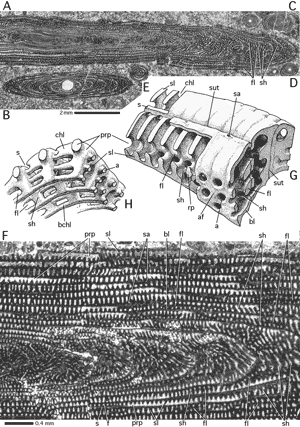
Figure 70: Odd association and polar structures in Praealveolina tenuis . From Alcantara, Lisbon; Cenomanian. Transmitted light micrographs.
A: microspheric specimen, axial section; B: megalospheric specimen, axial section; C-E: odd partners: C: axial section of microspheric Simplalveolina sp.; D: axial sections of megalospheric Simplalveolina sp.; E: Ovalveolina sp, axial section. F: microspheric P. tenuis, axial section, detail showing polar structure with floors and shaftsin the basement.
G: model of two subsequent chambers near their polar ends where the first floors in the basement appear. H: model of the protoplasmic body filling the cavities in G. Both models schematic, not to scale, after , 1933.
a: aperture: af: apertural face; bchl: basement chamberlets; bl: basal layer; chl: (main) chamberlets; fl: floor; prp: preseptal passage; rp: (incipient) residual pillar; s: septum; sa: supplementary aperture; sh: shaft; sl: septulum; sut: cameral suture.
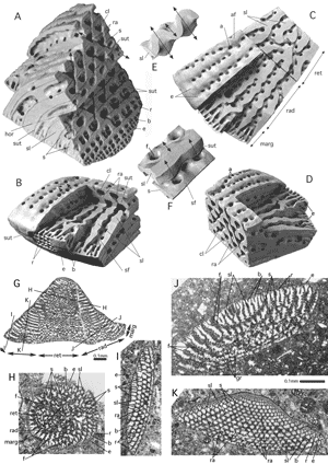
Figure 71: The structure of Orbitolina.
A-D: Oblique and vertical views of the cone base. These plasticine models
sculptured in the years around 1955 by (* 1896 - † 1984) were never published. E-F: Details of a septulum in the radial zone of the discoidal chamber. In model E the septulum is cut below the roof and above the bottom of the chambers. The apparent folding of the section results from the adjustment of the endoskeletal structure to the crosswise-oblique arrangement of the stolon axes (arrows) that produce so-called ramps. In model F the septulum is cut in the middle of the chamber and shows a part of the chamber bottom with the face of the previous chamber. In the middle of the chamber, the section of the septulum appears unfolded. Compare Fig. 47H and Fig. 80G. G-H: Random thinsections of Orbitolina sp. from Southwestern France, Albian. Transmitted light micrographs. In Fig. G the approximate positions of sections H-K are indicated. Note the inverse orientation: the sections face downward. G-K: Random sections of Orbitolina sp. from Southwestern France, Albian. Transmitted light micrographs. The approximate position of sections H-K are indicated in section G that is very close to the axial plane.
For 's zonation of the chamber see Fig. 20. Section H demonstrates details of the reticular zone, section I details of the marginal zone and section J details of the radial zone. The transverse section K shows the ramps produced by
the crosswise-oblique stolon system.
a: aperture; af: apertural face; b: beam; cl: chamberlet; e: epiderm; f: foramen; gr: coarse grains in the septum that obscure the structural pattern; hor: horizontal section in the plasticine model; marg: marginal zone; r: rafter; ra: ramp; rad: radial zone; ret: reticular zone; s: septum; sf: septal face; sl: septulum; sut: suture of the chambers. Double arrows in E and F: crosswise oblique foraminal axes.
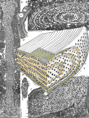
Figure 72: The structure of Orbitopsella:
a simple exoskeleton and a pillared endoskeleton in a discoidal shell: Orbitopsella dubari , Bou Dahar, Eastern Morocco, Middle Lias. Transmitted light micrographs.
A: oblique section of complete microspheric specimen; B: oblique section of microspheric specimen. The septa of the thickened margin are cut tangentially and reveal the alternating pattern
in the disposition of foramina on the septal face. C: oblique tangential section of a microspheric specimen at a low angle to the equatorial plane showing a part of
the disc with its exoskeleton (restricted to beams). Note the large open spaces, the lateral annular passages (arrows), that
separate exoskeleton and endoskeleton. D: Oblique centered section of megalospheric specimen. Note the structured wall of the embryo, that shows it to be a sphaeroconch. E: Transverse section (parallel to
the axis of coiling) of a megalospheric specimen. The septum in this tangential section reveals the alternating pattern of the apertures. F: schematic model of structure after , 1967; not to scale.
Green: exoskeleton; brown: endoskeleton.
a: aperture; ap: annular passage; b: beam; f: foramen; p: pillar; pr: sphaeroconch; s: septum.
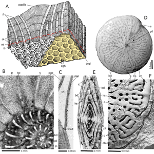
Figure 73: Papillae, beads and axial piles of lamellae.
A: papilla and pores. Stereograph, schematic, not to scale; after , 1977. B-C: Assilina madagascariensis (d') from Mauritius. Recent. Transverse section parallel to equatorial plane (B) and axial section (C) with minute papillae on
the last whorl; transmitted light micrograph. D-F: Beading in Amphistegina papillosa . Gulf of Aqaba, Red Sea; Recent. D: dorsal view of shell showing beading of septular and hemiseptular sutures. Incident light micrograph. E: axial section showing hemiseptular and septular support of beads; transmitted light micrograph.
F: Epoxy resin cast of shell cavity showing spiral main chamber lumina in dorsal view, interrupted by the hemiseptula supporting the interseptal beads, SEM graph.
a: aperture; bd: beads (septular and hemiseptular); ch: main (spiral) chamber lumen; egh: eggholders; hs: hemiseptulum; il: inner lamella; isc: intraseptal canal system; ml: median layer; ol: outer lamellae; orgl: organic lining of protoplast; p: pores; pap: papillae; pil: axial pile of lamellae; s: septum and septal suture; wsut: whorl suture.
Figure 74: Parachomata in Pseudodoliolina sp. from a commercial
thin section labeled "Moscow", Permian.
A: oblique section at low angle to axis of coiling (arrow). B: oblique section nearly perpendicular to the axis of coiling.
c*: choma; f: foramina forming a single row in alternation with the parachomata; s: septum (without fluting); spth: spirotheca (no trace of keriotheca).
The magnification of each figure is in proportion to the length of the 10 cm scale bar above.
Figure 75: The pore and its organic constituents.
A: a pore in an inner chamber covered by an outer whorl, according to (1977), schematic, not to scale. B: Accumulation of mitochondria below a pore mouth in Bolivina sp.,
thus indicating the pores' main function: gas exchange. TEM micrograph of a section oblique to the surface ot the wall that
exaggerates the thickness of the pore discs. The detachment of the outer lamella 2 (ouL 2) is an
artifact of preparation. x 24,000. Courtesy . C: Resin cast of pores in
the lateral chamber wall of Nummulites partschi with trabeculae. The carbonates of the shell are dissolved with HCl. SEM graph x 1,000. D: Outer pore mouths in
the lateral suface of chamber wall of Assilina. Note the annular attachment of the interlamellar discs. SEM graph x 5,000. E: Inner pore mouths in the lateral chamber wall of Assilina shaped as eggholders (in order to keep the symbionts below their breathing
chimneys). Note the annular suture of the pore plug. SEM graph x 5,000. C-E from , 1977. F: Perforation pattern on the dorsal surface of Challengerella persica (Recent, Persian Gulf): densely perforated porefields between imperforate ornamentation. SEM: oblique dorsal view of shell, x 30, and detail of porefield, x 500. G: Perforation pattern in Ammonia reyi (Pliocene, Dar bel Hamri, Northern Morocco): densely perforated porefields between loosely perforated ornaments. SEM graphs of dorsal shell view (x 30) with
detail (x 500). F-G: from et alii, 1980.
Abbreviations: bD: basal (pore) disc; Cy: cytoplasm; iL: inner lamella; ilD: interlamellar disc; iol: interlamellar organic lining; ls: lacunar system (in the cytoplasm); M: mitochondria; ML: median layer separating inner from outer lamellas; OL: Organic lining (here difficult to separate from plasmalemma of host); ouL 1: primary outer lamella; ouL 2, 3: subsequent outer lamellas; P: pore; Pc: organic pore coat; Pl: plasmalemma; ppl: pore plug (note its porosity); Spl: (biomineralized) sieveplate; V: vacuole.
Figure 76: A: selliform deformation of the lenticular-compressed test
of Eulepidina sp., Oligocene, Sarawak, Borneo. Transverse section approximately perpendicular to the axis of the shell. Transmitted light micrograph. B: sketch of a selliform shell with its axis. Schema, not to scale.
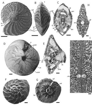
Figure 77: Umbos and umbilical plugs.
A-C: Elphidium craticulatum ( et ) from the Gulf of Aqaba, Red Sea. Recent. A: lateral view, SEM graph. B: peripheral view, SEM graph. C: slightly oblique axial section cutting through the symmetrical pair of umbilical plugs. Note the presence of spaces in the biumbilical shell,
caused by spiral canals and funnels. Transmitted light micrograph. D-E: Amphistegina lessonii d' from the Gulf of Aqaba, Red Sea. Recent. D: ventral view shows the stellar chamberlets overgrowing
part of the ventral umbo. Incident light micrograph. E: axial section of specimen having lost the last few chambers. There is a smaller dorsal and a larger ventral umbo. F-G: Ammonia umbonata (). Dorsal and ventral views, SEM graphs. The ventral (umbilical) view exhibits a free-standing, composite umbilical plug. H-I: the umbos in porcelaneous shells may react to diagenetic processes by
differences in recrystallisation that possibly indicate a differentiation in the
texture of the wall in the umbonal area of the shell. Diagnostic for the involute stages
in the growth of Meandropsinidae in the Upper Cretaceous of the Pyrenean Gulf. H: megalospheric Fascispira sp. from Canelles, Lerida, Northern Spain. Axial section. I: microspheric Larrazetia larrazeti (), center of axial section, from Bac de Grillera, Northern Spain. All scale bars 0.1 mm.
a: aperture; af: apertural face; cpl: coverplate; f: foramen; fo: fossette; fpl: foramenal plate; fu: funnel; k: keel; m: mask; pust: pustules on amphisteginid face; rp: retral process; s: septum; spc: spiral canal; ub: umbo; up: umbilical plate; upl: umbilical plug.
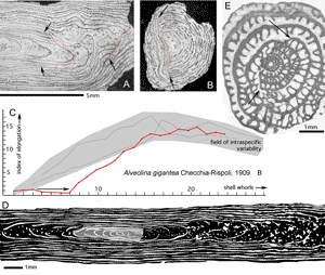
Figure 78: Regeneration in fusiform-elongate shells: A-D: Alveolina gigantea . Microspheric generation. Palermo, Sicily, Middle Eocene. E: Pseudoschwagerina sp., megalospheric specimen, from Palazzo Adriano, Sicily, Permian.
A: a fragment of the polar realm of the shell cut in the axial direction shows two periods of regeneration (red lines designated by arrows) during late ontogeny. B: a similar fragment cut in
a direction perpendicular to the shell axis, with one broken surface, that has been regenerated in an attempt to restore the cylindrical shape of the shell. At
such a late ontogenetic stage the original shape of the shell can not be regained through regenerative growth. Incident light micrographs. C-D: regeneration at an early stage of ontogeny:
C. the rate of elongation was higher in the regenerated half of the shell (red curve) than in the undisturbed half, until the range of elongation
that is characteristic for the species was attained again. Note the absence of elongation index increase in the early whorls broken away (arrow). Data from , 1962.
D. Compare the camera lucida drawing of an axial section: broken early shell in gray, regenerated shell in black. E: in a fusulinid the regeneration of the broken whorls 3-6 produces an almost perfect equatorial spiral after only two more whorls of growth.
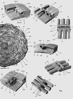
Figure 79: Preseptal (prp) and postseptal (pop) passages.
A: Alveolina munieri (equatorial section of microspheric specimen, Middle Lutetian, S. Giovanni Ilarione, Northern Italy).
B-E: unpublished plasticine models of the shell by . B-C: oblique views
showing chamberlets. In C the chamber roof (spirotheca) is partly removed. D: side view of chamber showing septum and passages behind and
in front of it. E: oblique view complementary to B showing main and intercalary apertures. the septal realm between two adjacent chambers. F-H:
unpublished models of sarcode (=shell cavities) by seen from above (F), obliquely from the side (G) and from below (H); Arrows: direction of growth.
bl: basal layer; chl: chamberlet; chlsut: chamberlet suture; chsut: chamber suture; f: foramen; icf: intercalary foramen; pop: postseptal passage; prp: preseptal passage; prwsurf: chamber roof surface of previous whorl; s: septum; sl: septulum; sup: supplementary passages in basal layer; sut: suture (of chambers); te: chamber roof (tectum).
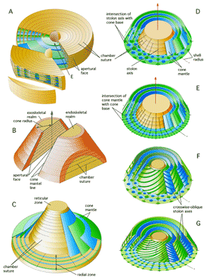
Figure 80: Stolon planes and foramenal axes in discoidal-annular and conical-uniserial shells. Schematic, not to scale.
A: Discoidal shell with a broadening periphery. Green and blue stolon planes are added step by step to the equatorial plane (E) as the shell margin thickens during ontogeny. The sector cut from the disc is cut in
its turn in a transverse direction. B: A cone composed of a single series of discoidal chambers
of which the marginal and axial areas are differentiated by colour. Note the distribution of the marginal area, the emplacement of the exoskeleton, and of the axial area housing the endoskeleton, in axial, horizontal (basal) and transverse sections. The axis
of the shell is indicated by a vertical arrow. The surface of the cone is called
the cone mantle, its horizontal termination is the cone base. A vertical line on the
slanting surface of the cone is called a cone mantle line. The cone radius is indicated by a double arrow. C: The stolon axes are distributed on cone mantles
in conical- uniserial foraminifera. If the cones increase their radial dimension
markedly during growth, additional cone mantles are added in order to maintain the radial distances between the cone mantles
relatively constant. This addition of cone mantles disturbes the regularity of the endoskeletal structures. In the models D-G, the addition of cone mantles during ontogeny is not taken into account. D-G: arrangement of stolon axes on cone mantles in the so-called radial zone of the cone (Fig. 20)
is in accordance with the four basic patterns that govern discoidal structures. In conical shells, however, the stolon planes are
replaced by cone mantles. As the cone increases in radius during growth, new stolon axes are intercalated
in the cone mantles (arrows). D: stolon axes in the cone mantles alternate in radial position
as a mantle is added, e.g. Dictyoconus. E: arrangement of stolon axes on cone mantle lines aligned on a cone radius. F: crosswise-oblique arrangement of stolon axes
alternating in radial position on successive cone mantles. G: crosswise-oblique arrangement of stolon axes
in line on a shell radius on subsequent cone mantles. This structure is a characteristic
of Orbitolina (Fig. 71).
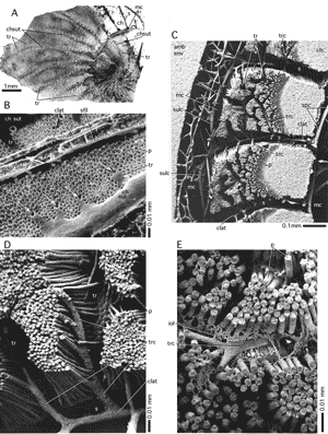
Figure 81: Trabeculae.
A: trabecules in a totally evolute nummulite, Nummulites giganteus (). Crimea. Lower Eocene (Cuisian). Transmitted light micrograph of a section tangential to the lateral chamber walls. B-D:
unfilled shells of Nummulites planulatus () from Bos d'Arros, Gan, Southwestern France, Cuisian. B: lateral surface of the shell with a part of a septal suture between alar prolongations and their extensions, the trabeculae. The white arrows indicate the orifices of the trabecular canals. The lateral surface is overgrown by the next whorl
that has been broken away above the basal suture of a septum of an alar prolongation leaving a
linear mark called septal filament. At the base of the septum of an alar prolongation there is a single intraseptal canal running
the lenght of the alar prolongation. Where such a canal crosses a trabecular orifice (arrow), a passage may be created by partial resorption of the wall. This passage connects canal cavities of two successive whorls. SEM graph. C: epoxy-resin cast of shell cavities cut in
an approximately equatorial direction showing two complete chambers with their marginal cord. Note the "grooves" in the porous wall created by the trabecular canals. SEM graph. D: detail of C showing the deviation of the pores in order to admit a trabecular canal. E: trabecular canal in Nummulites partschi from Bos d'Arros, Gan. Cuisian. Epoxy resin cast. The preservation of the interlamellar organic lining as
a cavity in the shell permits the oblique path of the trabecular canal to be followed through subsequent lamellae.
Abreviations: amb env: ambient environment appearing in a cast of cavities as solid mass; ch: chamber lumen; chsut: chamber suture; clat: lateral intraseptal canals in the chamber and in the alar prolongations; iol: interlamellar organic layer; mc: marginal cord; p: pores; sfil: septal filament; spc: spiral canal; sulc: sulcus; tr: trabecule; trc: trabecular canal.
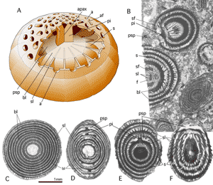
Figure 82: Trematophores.
A: stereograph of the trematophore of Lacazinella supported by a
single residual pillar in axial position. Schema, not to scale. B-F: Lacazinella wichmanni () from the Vogelkop, New Guinea. Middle-Upper Eocene. Transmitted light micrographs. All specimens are megalospheric. B: sections tangential to a septal face. C:
a section perpendicular to the apertural axis reveals the concentric growth of the chambers from their
beginning. D: section in the apertural axis. Note the alternation of the trematophore on opposite poles of the shell in successive chambers. E: oblique section cutting the apertural axis in a pillar supporting the trematophore. F: oblique section not cutting the apertural axis within the shell. Note the interruption of the septula below the equatorial roof of the chambers.
a: aperture; af: apertural face; apax: apertural axis; bl: basal layer; f: foramen; pi: pillar; psp: preseptal space; s: septum by transformation of the outer chamber wall that is totally covered by a new concentric chamber; sf: septal face; sl: septulum.
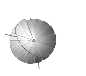
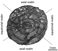
Figure 83: Model in polar view and a globular Alveolina
(Middle (?) Eocene of Pakistan) cut in a similar direction. Note the shortened
distance between the septa in the equatorial and the lengthened distance in the
axial realm. In oblique sections of globular to elongate involute planispiral shells the structure of the chambers in opposite sectors of the section approaches more or less closely in appearance that of either an equatorial or an axial section depending on the location and angle of the cut with respect to the septa.